1. Overview
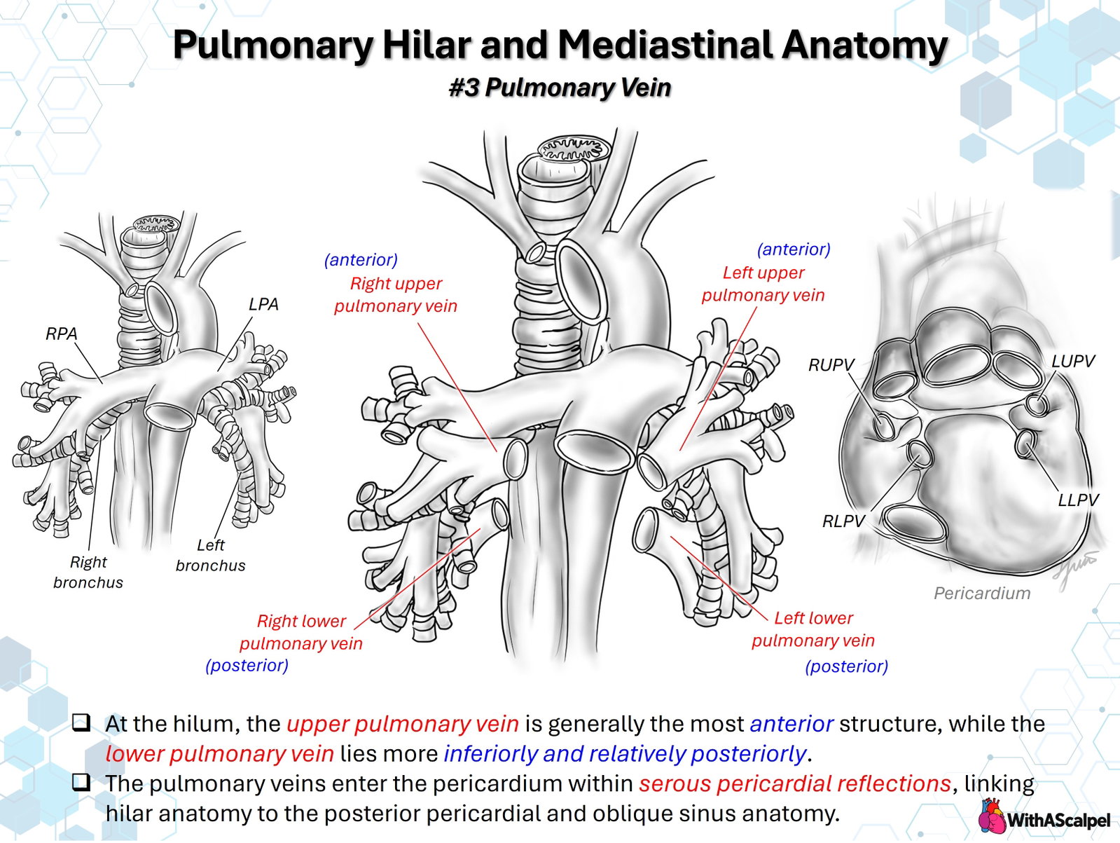
The pulmonary veins connect the pulmonary microcirculation to the left atrium and form an essential component of each lung root. Their organization is best understood at two different levels: the hilum, where venous branches pass among bronchi and pulmonary arteries, and the venoatrial junction, where the veins traverse pericardial reflections and enter the posterior left atrium. The conventional arrangement consists of a superior and an inferior pulmonary vein on each side. At either hilum, the superior pulmonary vein is generally the most anterior major structure; the inferior pulmonary vein lies more caudally and relatively posteriorly. These are useful spatial relationships rather than invariant rules for every venous branch [1,2].
This distinction matters in cross-sectional imaging, lung resection, pulmonary venous reconstruction, and operations involving the posterior left atrium. A schematic anterior view conveys the overall configuration, but safe interpretation requires attention to depth, lobar tributaries, venous ostia, and the pericardial boundary.
2. Pulmonary Venous Organization at the Hilum
Pulmonary veins collect oxygenated blood from intrapulmonary venules and converge through segmental and lobar tributaries. Unlike the pulmonary arteries, which generally accompany segmental bronchi, many pulmonary venous tributaries follow intersegmental planes. This separation helps define anatomical pulmonary segments during surgery. Near the lung root, the tributaries form larger veins that run toward the left atrium, usually anterior and inferior to the principal pulmonary arterial and bronchial structures [1].
In the classic four-vein configuration, the right superior pulmonary vein (RSPV) drains the right upper and middle lobes, while the right inferior pulmonary vein (RIPV) receives most right lower-lobe drainage. The left superior pulmonary vein (LSPV) collects blood from the left upper lobe, including the lingula, and the left inferior pulmonary vein (LIPV) drains the left lower lobe. This arrangement describes the predominant territories, not a guarantee that all segmental veins join the expected trunk [3].
The anterior–posterior relationship changes between the peripheral hilum and the medial venoatrial junction. A vein that looks anterior to a bronchus in the lung root may turn posteriorly as it approaches the left atrium. Furthermore, the superior and inferior pulmonary venous orifices are offset vertically and in depth. Their position must therefore be assessed in axial, sagittal, and coronal planes rather than inferred from a single projected drawing.
3. Right Pulmonary Veins
3.1 Right superior pulmonary vein
The RSPV courses medially from the anterior right hilum toward the upper right aspect of the posterior left atrial wall. It generally lies anterior and inferior to the right pulmonary artery at the hilum and anterior to the central bronchial pathway. The proximal right pulmonary artery itself passes behind the ascending aorta and superior vena cava (SVC), so the apparent arterial–venous relationship depends on where the vessels are examined.
The RSPV usually receives upper-lobe segmental drainage and a tributary from the middle lobe. The latter may join the RSPV, enter the left atrium independently, or join an inferior venous channel. Close to the atrium, the RSPV is related to the posterior aspect of the SVC and the right atrial–left atrial interface. These landmarks are important when distinguishing a normally connected RSPV from an anomalous connection to the SVC or right atrium [3].
3.2 Right inferior pulmonary vein
The RIPV forms from the superior segmental and basal venous tributaries of the right lower lobe. It approaches the inferoposterior right atrial aspect of the left atrium and is commonly deeper than the RSPV. Its proximal tributaries may divide very close to the atrial orifice, leaving only a short segment available for mobilization or anastomosis.
The inferior pulmonary ligament is a caudal pleural continuation of the lung root, below the inferior vein. It is a useful thoracic surgical landmark, but the ligament is not a pulmonary vein and should not be confused with the serous pericardial reflections surrounding the intrapericardial venous segments. On the right, the RIPV also lies close to the pericardial reflection extending toward the inferior vena cava.
4. Left Pulmonary Veins
4.1 Left superior pulmonary vein
The LSPV emerges from the anterior left upper-lobe hilum and courses medially toward the superior left atrial wall. It usually collects venous drainage from the upper-lobe segments and lingula. At the lung root, it lies predominantly anterior to the bronchial structures, while the left pulmonary artery passes superior to the left main bronchus and continues into the lung.
Near the heart, the LSPV is closely related to the left atrial appendage, which lies anterior and somewhat lateral to its venoatrial junction. The intervening tissue ridge is important in left atrial anatomical orientation and electrophysiologic procedures [2]. In cardiac surgery, the juxtaposition of the appendage and superior vein also helps orient the lateral left atrial wall.
4.2 Left inferior pulmonary vein
The LIPV receives the superior and basal venous tributaries of the left lower lobe. It lies inferior to the LSPV and usually courses more posteriorly as it approaches the left atrium. The descending thoracic aorta is a relevant posterior landmark, and the left lower-lobe bronchus lies nearby laterally.
The left inferior venous ostium typically occupies a more posterior and inferior position than the LSPV ostium. In a posterior left atrial view, the distance and intervening atrial wall between these orifices can vary substantially. A conjoined left pulmonary venous trunk may replace the two distinct venous channels depicted in the conventional illustration [3].
5. Left Atrial Junction and Pericardial Reflections
The pulmonary veins do not simply terminate at the outer surface of the pericardium. They pass from the lung roots through serous pericardial reflections to reach the left atrium. The proximal venous segments and the venoatrial junctions therefore have an intrapericardial relationship, while more peripheral veins are surrounded by hilar connective tissue and pleura. The length and configuration of this transition vary among patients.
The posterior left atrium forms the central surface around which the veins enter. At the junction, atrial myocardium extends variably onto the pulmonary veins as myocardial sleeves. A human anatomical study demonstrated more developed venous myocardial sleeves and junctional muscle formations around superior than inferior veins, although individual variation is substantial [4]. These sleeves explain the electrophysiological importance of the pulmonary venous antra and also reinforce that the venoatrial junction is not a simple, uniformly cylindrical tube.
The serous pericardium reflects around the pulmonary veins and adjacent caval structures, creating recesses rather than one uninterrupted free posterior pericardial surface. In a study of 107 cadaveric dissections, investigators documented variation in pulmonary venous pericardial segments and classified several configurations of the posterior pericardial reflections [5]. Thus, the attachment of the pericardium should be identified in the patient rather than assumed from a standard illustration.
5.1 The oblique pericardial sinus
The oblique sinus is a blind pericardial recess behind the left atrium. Its lateral boundaries are formed largely by the reflections around the right and left pulmonary venous complexes, and its inferior boundary is related to the reflection around the inferior vena cava. Its posterior surface faces the esophagus and other posterior mediastinal structures across the pericardial tissues.
From the anterior operative field, elevation of the heart and careful entry behind the left atrium allow access to this recess. The oblique sinus provides an orientation for posterior left atrial exposure and some pulmonary venous reconstructions. It must not be confused with the transverse sinus, which lies superior and anterior to the left atrium, behind the arterial great vessels.
The phrenic nerves travel on the external surface of the fibrous pericardium, anterior to the respective lung roots. They are therefore important during lateral pericardial opening and extensive hilar mobilization, even though they are not part of the serous venous reflections themselves.
6. Developmental Basis
The pulmonary venous component of the left atrium forms through development of the common pulmonary venous connection and incorporation of its branches into the expanding atrial wall. The mature pattern of four separate pulmonary venous ostia arises through this remodeling rather than from four independent primitive veins.
High-resolution reconstructions of 23 human embryos and 19 fetuses demonstrated developmental changes in the pulmonary veins, left atrium, appendage, and pericardial relationships [6]. These observations connect the mature venous ostia with the remodeling of the posterior atrial wall and pericardial attachments. Abnormal incorporation or persistence of embryonic venous connections can produce anomalous pulmonary venous drainage; the developmental explanation is useful, but the actual course of an anomaly must still be defined by imaging.
7. Normal Variants and Their Significance
Four separate ostia are common but not universal. Variations include a conjoined superior–inferior trunk, a separate middle-lobe or accessory vein, and early division into multiple tributaries near the atrium. An ostial variation should be distinguished from anomalous pulmonary venous connection: the former still drains into the left atrium, whereas the latter directs pulmonary venous blood to a systemic venous structure or the right atrium [3].
CT studies and a systematic review establish substantial diversity in pulmonary venous ostial anatomy [7,8]. In one preoperative three-dimensional CT series of 140 patients, common left-sided ostia were reported in 33%, and a separately draining right middle-lobe vein in 8% [9]. In another CT series of 135 patients, a long common left trunk occurred in 11.1%, while independent middle-lobe drainage was identified in approximately 25% [10]. These figures are study-specific, reflecting differences in populations and anatomical definitions; they should not be combined into one universal prevalence estimate.
An especially important distinction is between a true common trunk and two adjacent separate ostia with little intervening atrial wall. Likewise, an early branching vein is different from an accessory vein entering the atrium through an independent orifice. Such details affect safe venous division during lung resection, planning of catheter ablation, and the feasibility and geometry of pulmonary venous reconstruction.
8. Imaging and Three-Dimensional Assessment
Transthoracic and transesophageal echocardiography can identify pulmonary venous drainage into the left atrium, Doppler flow abnormalities, and associated cardiac lesions. However, a satisfactory view of one or two veins does not establish the anatomy of all pulmonary venous channels. The more peripheral or posterior portions may be incompletely visualized, especially in complex congenital anatomy or after prior surgery.
CT angiography with multiplanar and three-dimensional reconstruction is particularly useful for mapping the number of ostia, individual tributaries, relationship to bronchi and pulmonary arteries, and the position of the veins relative to the SVC, left atrium, and descending aorta. Modern imaging reviews emphasize reporting the venous course as well as the site of drainage, since an apparently normal atrial orifice does not exclude important upstream branching or narrowing [11].
When pulmonary venous intervention or anomalous venous repair is contemplated, a practical anatomical assessment identifies: (1) each lung segment's venous drainage; (2) all venoatrial or anomalous connections; (3) any common trunk or early bifurcation; (4) the relationship to the airway and arterial tree; and (5) any focal stenosis, compression, or hypoplastic segment. In congenital anomalous pulmonary venous connection, CT can clarify connections and adjacent vascular structures that are difficult to define completely by echocardiography [12]. MRI is an alternative when flow quantification and avoidance of ionizing radiation are priorities, provided spatial and temporal resolution are adequate.
9. Procedural and Surgical Implications
For lung resection, the anterior superior pulmonary vein is commonly encountered early during hilar dissection. An independent middle-lobe vein or an unexpected interlobar drainage pathway must be recognized before vascular division. Injury to an unrecognized tributary can cause hemorrhage; division of a vein that drains retained lung can produce venous congestion. Preoperative venous mapping becomes particularly important for segmentectomy, where preservation of intersegmental venous drainage determines the functional anatomy of the remaining lung [10].
For cardiac surgery, the transition from extrapericardial hilar vein to intrapericardial venous segment determines the available mobilization plane. Dividing pericardial attachments can provide exposure, but it does not eliminate fixation from distal tributaries, bronchovascular connective tissue, or short venous branches. Excessive traction, torsion, or a narrow anastomotic angle may impair venous return even when the final suture line appears widely patent.
For pulmonary venous reconstruction, the surgeon must distinguish the true venoatrial junction from a common vein and any adjacent early branches. The objective is an unobstructed drainage pathway without circumferential distortion or tension. In repairs of anomalous pulmonary venous connection, the relationship between the right-sided veins, SVC, and cavoatrial junction may determine whether an intracardiac baffle or caval reconstruction is anatomically appropriate [12].
The final assessment should focus on all pulmonary venous pathways, not simply the reconstructed or most visible vein. Echocardiography or other imaging should evaluate residual gradients, flow acceleration, atrial inflow geometry, and potential compression. These are procedure-specific applications of the anatomy rather than a recommendation for one universal reconstructive technique.
Key Clinical Principles
- Superior vein: usually the most anterior major structure at either hilum.
- Inferior vein: more caudal and often relatively posterior, but not necessarily a single long trunk.
- Venoatrial junction: defined by venous entry into the posterior left atrium and variable myocardial sleeves.
- Pericardial reflections: surround proximal pulmonary venous segments and establish the lateral boundaries of the oblique sinus.
- Variant drainage: common trunks, accessory ostia, and early branches must be distinguished from anomalous systemic venous connections.
- Operative imaging: follow each pulmonary vein from its lobar tributaries to its final atrial or systemic venous destination.
https://x.com/WithAScalpel/status/2124125864694829377?a=n00BrJR2QoPjiBohSCI&s=20
References
- Kandathil A, Chamarthy M. Pulmonary vascular anatomy & anatomical variants. Cardiovasc Diagn Ther. 2018;8(3):201–207. DOI: 10.21037/cdt.2018.01.04. PMID: 30057869.
- Ho SY, Cabrera JA, Sánchez-Quintana D. Left atrial anatomy revisited. Circ Arrhythm Electrophysiol. 2012;5(1):220–228. DOI: 10.1161/CIRCEP.111.962720. PMID: 22334429.
- Pandey NN, Taxak A, Kumar S. Normal pulmonary venous anatomy and non-anomalous variations demonstrated on CT angiography: what the radiologist needs to know? Br J Radiol. 2020;93(1116):20200595. DOI: 10.1259/bjr.20200595. PMID: 32706997.
- Nathan H, Eliakim M. The junction between the left atrium and the pulmonary veins: an anatomic study of human hearts. Circulation. 1966;34(3):412–422. DOI: 10.1161/01.CIR.34.3.412. PMID: 5922708.
- Chaffanjon P, Brichon PY, Faure C, Favre JJ. Pericardial reflection around the venous aspect of the heart. Surg Radiol Anat. 1997;19(1):17–21. DOI: 10.1007/BF01627729. PMID: 9060112.
- Fukui N, Kanahashi T, Matsubayashi J, et al. Morphogenesis of the pulmonary vein and left atrial appendage in human embryos and early fetuses. J Anat. 2024;244(1):142–158. DOI: 10.1111/joa.13941. PMID: 37559438.
- Marom EM, Herndon JE, Kim YH, McAdams HP. Variations in pulmonary venous drainage to the left atrium: implications for radiofrequency ablation. Radiology. 2004;230(3):824–829. DOI: 10.1148/radiol.2303030315. PMID: 14739316.
- Bonczar M, Piątek-Koziej K, Wolska J, et al. Variations in human pulmonary vein ostia morphology: a systematic review with meta-analysis. Clin Anat. 2022;35(7):906–926. DOI: 10.1002/ca.23896. PMID: 35460116.
- Akiba T, Marushima H, Odaka M, et al. Pulmonary vein analysis using three-dimensional computed tomography angiography for thoracic surgery. Gen Thorac Cardiovasc Surg. 2010;58(7):331–335. DOI: 10.1007/s11748-010-0592-0. PMID: 20628848.
- Polaczek M, Szaro P, Jakubowska L, et al. Pulmonary veins variations with potential impact in thoracic surgery: a computed-tomography-based atlas. J Thorac Dis. 2020;12(3):383–393. DOI: 10.21037/jtd.2020.01.34. PMID: 32274104.
- Hassani C, Saremi F. Comprehensive cross-sectional imaging of the pulmonary veins. Radiographics. 2017;37(7):1928–1954. DOI: 10.1148/rg.2017170050. PMID: 29131765.
- Kasahara H, Aeba R, Tanami Y, Yozu R. Multislice computed tomography is useful for evaluating partial anomalous pulmonary venous connection. J Cardiothorac Surg. 2010;5:40. DOI: 10.1186/1749-8090-5-40. PMID: 20482753.