1. Overview
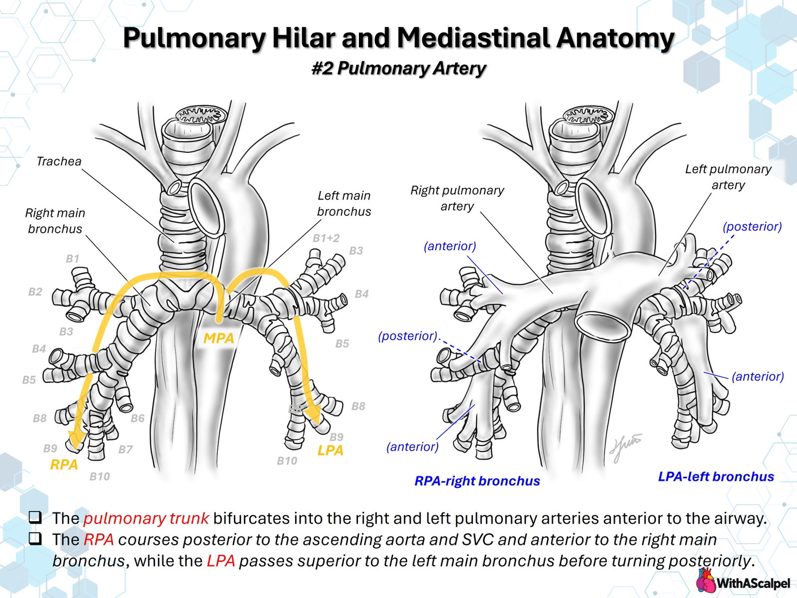
The pulmonary trunk and its branches form the vascular component of the pulmonary roots, closely interwoven with the tracheobronchial tree. Their course is asymmetric: the right pulmonary artery (RPA) travels posterior to the ascending aorta and superior vena cava (SVC) before crossing anterior to the right main bronchus, whereas the left pulmonary artery (LPA) arches superior to the left main bronchus and then follows a more posterior course toward the left lung. These relationships are fundamental to interpreting cross-sectional imaging, understanding extrinsic airway compression, and planning pulmonary arterial reconstruction.
A schematic anterior view demonstrates the basic relationships but cannot show every depth transition or segmental variation. The hilar anatomy should be considered in three dimensions, with attention to the point where an artery crosses a bronchus, the origin of its lobar branches, and the adjacent great vessels [1,2].
2. Pulmonary Trunk and Bifurcation
The main pulmonary artery (MPA) originates from the right ventricular outflow tract and courses superiorly, posteriorly, and toward the left in the usual ventriculoarterial arrangement. Its bifurcation lies anterior to the distal airway, generally inferior to the aortic arch and near the region of the tracheal bifurcation. From this junction, the RPA travels horizontally toward the right hilum, while the LPA follows a shorter, oblique, leftward course.
The spatial position of the bifurcation is influenced by the relationship between the MPA, ascending aorta, aortic arch, and carina. Although the central pulmonary arteries are commonly described as anterior to the airway, their distal pathways are not uniformly anterior: each branch changes direction as it approaches and enters its respective lung. The illustration's anterior and posterior labels therefore refer to particular artery–bronchus intersections, not to an invariant relationship along the complete pulmonary arterial tree.
The intrapericardial proximal pulmonary arteries are continuous with more distal extrapericardial vessels at the pericardial reflections surrounding the great vessels. This distinction matters when mobilization is required: gaining length centrally does not necessarily release distal fixation at the lung root, and excessive traction can deform a branch or its first lobar origins.
3. Right Pulmonary Artery: Mediastinal and Bronchial Relationships
The RPA is typically the longer of the two branch pulmonary arteries. Leaving the bifurcation, it passes posterior to the ascending aorta and SVC. This deep mediastinal course explains why the proximal RPA may be partially concealed in an anterior operative view and why its caliber can be difficult to assess from a single echocardiographic plane.
As the vessel reaches the right lung root, the RPA lies anterior to the right main bronchus. The relationship changes after the origin of the right upper lobe pulmonary arterial branches: the interlobar pulmonary artery proceeds inferiorly and laterally, closely associated with the bronchus intermedius and subsequently the lower-lobe airway branches. Accordingly, “RPA anterior to the right bronchus” is most useful as a proximal hilar orientation rather than as a description of every lobar artery.
The right upper lobe bronchus commonly arises above the pulmonary artery at the hilum. It is consequently termed the eparterial bronchus, in contrast to the middle- and lower-lobe bronchi, which arise below the level of the artery and are termed hyparterial. This configuration creates a distinctive right-sided hilar pattern and explains why the highest right hilar airway branch may appear superior to the adjacent pulmonary artery despite the artery lying anterior to the main bronchus [1–3].
The azygos arch, SVC, ascending aorta, and upper-lobe bronchovascular structures are useful landmarks. In operations involving the proximal RPA, dissection beneath or behind the ascending aorta must preserve awareness of both the posterior airway and the more anterior systemic venous and aortic structures.
4. Left Pulmonary Artery: Its Arch over the Left Main Bronchus
The LPA originates from the pulmonary bifurcation and courses leftward toward the lung beneath the aortic arch. Its defining hilar relationship is superior to the left main bronchus. Rather than passing horizontally in front of the bronchus throughout its length, the LPA curves over the airway and then follows a posterolateral or posterior trajectory as it gives rise to lobar and segmental branches.
The left main bronchus is longer and more obliquely oriented than its right-sided counterpart. Because no normal left upper lobe bronchus arises above the proximal LPA, the left lobar bronchi are described as hyparterial. In an anterior view, the vessel can appear to overlap the left main bronchus; sagittal and coronal reconstructions clarify that its proximal hilar course is superior rather than simply anterior.
The proximal LPA also has a close relationship with the ductus arteriosus or ligamentum arteriosum, which usually connects its proximal region to the aortic isthmus or undersurface of the arch. The left recurrent laryngeal nerve loops beneath the aortic arch near this attachment. These relationships are pertinent to ductal dissection, pulmonary artery mobilization, vascular ring surgery, and branch pulmonary artery reconstruction.
A markedly enlarged, displaced, or stented LPA can narrow the space available to the left main bronchus. Such compression is especially consequential when a prominent neoaortic root or altered postoperative great-vessel configuration creates an additional rigid boundary.
5. Lobar and Segmental Pulmonary Arterial Branching
5.1 Right lung
The first major RPA branch commonly supplies the right upper lobe as the truncus anterior (superior trunk). Additional ascending or recurrent branches to the upper lobe may arise from the interlobar artery. Distally, the interlobar RPA gives branches to the right middle lobe, the superior segment of the lower lobe (A6), and the basal segments (A7–A10).
Pulmonary arterial branches generally accompany the corresponding segmental bronchi. The numerical notation A1–A10 parallels the bronchial labels B1–B10, but the origins of the arteries are not necessarily identical to the bronchial branching points. The upper-lobe arterial supply is particularly variable, including separate, common-trunk, recurrent, and ascending patterns. CT angiographic and operative studies demonstrate that relying on a single presumed superior-trunk pattern risks overlooking accessory branches [5,6].
5.2 Left lung
The LPA generally provides branches to the apicoposterior region of the left upper lobe (frequently A1+2), the anterior segment (A3), and the lingula (A4 and A5). It then supplies the superior lower-lobe segment (A6) and basal segments, which often exhibit a combined anteromedial basal distribution rather than discrete A7 and A8 branches.
Left-sided upper-lobe and interlobar arterial branching is notably diverse. Lingular branches may originate at different levels, and posterior or interlobar branches may arise independently rather than through one stereotyped trunk. In a CT-based study of 320 patients, left interlobar branching patterns were classified into seven broad types, underscoring the need for patient-specific assessment before segmental intervention [7].
These descriptions represent common adult arrangements. They are useful for understanding the illustration's A/B labels, but should not be applied mechanically to congenital pulmonary vascular malformations or to surgically altered anatomy.
6. Hilar Orientation in Three Dimensions
A practical mnemonic is RALS: the right pulmonary artery lies anterior to the right main bronchus, while the left pulmonary artery lies superior to the left main bronchus. The mnemonic identifies an important asymmetric relationship, but it does not describe every artery–bronchus crossing within the lung.
The pulmonary veins form the third major component of the lung root and tend to occupy more anterior and inferior positions than the pulmonary arteries and central bronchi. Within the peripheral lung, segmental pulmonary arteries and bronchi generally travel together, whereas the pulmonary veins follow a more intersegmental course. Three-dimensional reconstruction studies show that these relationships can support anatomical segment identification and surgical navigation [1,3].
Hilar structures are not arranged in a rigid vertical stack. Cadaveric examination of 110 lung specimens identified anatomical variations in the numbers and positions of bronchi, pulmonary arteries, and veins, with greater observed variation on the left in that sample [4]. Such specimens illustrate anatomical diversity; their reported frequencies should not be assumed to apply directly to pediatric or congenital cohorts.
The mediastinal pleura invests the pulmonary root, and its inferior reflection continues as the pulmonary ligament. The phrenic nerve passes anterior to the lung root along the pericardium, while the vagus nerve typically passes posterior to it. These relationships help distinguish dissection planes during hilar mobilization, although the actual position of nerves and pleural attachments must be identified directly.
7. Congenital Variations and Airway Compression
The normal relationship between the pulmonary arteries and bronchi provides a reference for recognizing anomalous vessels. In a left pulmonary artery sling, the LPA typically originates from the RPA rather than directly from the MPA and travels posterior to the distal trachea, between the airway and esophagus, to reach the left lung. This is fundamentally different from the normal LPA passing above the left main bronchus.
Pulmonary artery sling may be accompanied by complete tracheal rings, fixed tracheal stenosis, abnormal bronchial branching, or pulmonary hypoplasia. In a CT series of 27 children with a sling, significant tracheobronchial narrowing was common, illustrating why defining arterial origin alone is insufficient [8]. Contemporary surgical series emphasize that the airway configuration, right lung development, degree of tracheal deviation, and available length for LPA reimplantation influence the operative strategy [9]. These findings come from selected patients with severe anomalies and should not be generalized to an isolated, asymptomatic vascular variant.
Acquired or postoperative airway compression can result from aneurysmal pulmonary arterial dilation, altered branch orientation, conduits, or implanted stents. The airway is particularly vulnerable when trapped between an expanded pulmonary artery and another relatively fixed vascular or mediastinal structure. Bronchomalacia may coexist with external narrowing, and an anatomically successful vascular procedure may not immediately reverse dynamic airway collapse.
In a retrospective series of 19 single-ventricle patients undergoing branch pulmonary artery intervention, two developed near-occlusion of the left main bronchus after stenting. The investigators subsequently emphasized cross-sectional assessment and selective concurrent bronchoscopy during intervention [10]. A separate cohort of 49 Fontan patients found an association between LPA stenting and smaller relative left main bronchial area, but could not establish whether the differences were caused by stent placement or preceded it [11]. These studies establish an important potential complication, not a universal contraindication to pulmonary artery stenting.
8. Imaging and Diagnostic Assessment
Echocardiography identifies the MPA bifurcation, proximal branch origins, flow acceleration, and associated cardiac lesions, but acoustic windows may not fully demonstrate the RPA behind the ascending aorta or the distal LPA beneath the arch. Doppler gradients also depend on flow and cannot, by themselves, define the extent of anatomical stenosis or the relationship to a bronchus.
CT angiography provides a direct three-dimensional map of the pulmonary arterial tree, airway lumen, aorta, and systemic veins. Review should extend beyond a volume-rendered image to thin axial source sections, multiplanar reconstructions, and curved reformations along the vessel and bronchus. CT-based comparisons of pulmonary arterial branches demonstrate that small vessels can be missed on three-dimensional renderings despite being detectable on thin-section multiplanar images [6].
For reconstruction or intervention, the imaging assessment should document the precise branch origin, stenotic length, reference-vessel diameter, first lobar branch points, arterial angulation, and contact with the bronchi. In airway-related cases, describe the site and length of narrowing, direction of compression, and any fixed tracheal stenosis. Cardiac MRI can supplement evaluation of branch pulmonary arterial flow distribution and ventricular function, although spatial resolution, respiratory motion, and the need to evaluate a small airway may favor CT in selected children.
Bronchoscopy adds information unavailable from static vascular imaging: pulsatile indentation, dynamic collapse, intrinsic stenosis, and changes during respiration or airway-pressure manipulation. Its results must be interpreted with the sedation conditions and ventilatory support in mind. Pulmonary artery angiography is valuable when intervention is being contemplated, but angiographic lumen measurements should be integrated with the airway assessment.
9. Implications for Surgery and Catheter Intervention
Branch pulmonary artery surgery requires more than an unobstructed luminal anastomosis. An effective reconstruction must preserve vessel length, orientation, and the neighboring airway. Before arterioplasty or reimplantation, define where the proximal vessel is fixed, where the first lobar branches arise, and whether the proposed patch or new course will create a sharp angle or anterior-to-posterior mass effect.
For the RPA, the ascending aorta and SVC can limit proximal exposure, and the right main bronchus lies immediately posterior to the hilar artery. For the LPA, mobilization beneath the arch must account for the ligamentum arteriosum, recurrent laryngeal nerve region, and the left main bronchus. Patch enlargement that improves the angiographic diameter may still leave a compressed airway or produce distortion at the distal end of the repair.
During catheter intervention, anticipated stent expansion should be assessed against the available space around the adjacent bronchus. In particularly high-risk anatomy, combined catheterization and bronchoscopy may reveal clinically important compression while the intervention is being performed [10]. Selected surgical rescue approaches, including pulmonary arterial translocation, have been reported for severe bronchial compression, but the evidence is limited to small series and individual cases [12].
Completion assessment should examine both vascular patency and bronchial geometry. Echocardiography or angiography evaluates residual gradients, flow distribution, and branch distortion; bronchoscopy or cross-sectional imaging may be necessary when ventilation remains asymmetric or airway obstruction is suspected. Persistent postoperative atelectasis, wheezing, focal air trapping, or extubation difficulty should prompt consideration of residual vascular compression, intrinsic stenosis, and bronchomalacia rather than an assumption that the pulmonary artery repair has addressed every cause.
10. Key Clinical and Surgical Principles
- Use RALS as an orientation, not a complete map: the proximal RPA is anterior to the right main bronchus; the LPA passes superior to the left main bronchus.
- Track each artery through its depth changes: the RPA runs behind the ascending aorta and SVC, while the LPA curves beneath the arch and over the airway.
- Identify lobar branch origins before reconstruction: segmental arterial patterns vary, particularly in the upper lobes.
- Evaluate the airway together with the artery: relief of pulmonary artery stenosis may coexist with new or persistent bronchial compression.
- Reconstruct geometry, not merely diameter: vessel angulation, fixation, branch takeoff, and airway clearance influence functional results.
- Interpret imaging in multiple planes: a visually convincing three-dimensional rendering must be checked against thin-section source images.
References
- Kandathil A, Chamarthy M. Pulmonary vascular anatomy & anatomical variants. Cardiovasc Diagn Ther. 2018;8(3):201–207. DOI: 10.21037/cdt.2018.01.04. PMID: 30057869.
- van der Spuy JC. The surgical anatomy of the pulmonary vessels. Thorax. 1953;8(3):189–194. DOI: 10.1136/thx.8.3.189. PMID: 13102416.
- Onuki T, Kanzaki M, Kikkawa T, et al. New findings on the three-dimensional anatomical relations between the bronchi and pulmonary blood vessels at the pulmonary hilum. Clin Anat. 2015;28(4):506–511. DOI: 10.1002/ca.22486. PMID: 25546314.
- Murlimanju BV, Massand A, Madhyastha S, et al. Anatomical variations of the arrangement of structures at the pulmonary hilum: a cadaveric study. Surg Radiol Anat. 2017;39(1):51–56. DOI: 10.1007/s00276-016-1716-8. PMID: 27338937.
- Fourdrain A, de Dominicis F, Blanchard C, et al. Three-dimensional CT angiography of anatomic variations in the pulmonary arterial tree. Surg Radiol Anat. 2018;40(1):45–53. DOI: 10.1007/s00276-017-1914-z. PMID: 28856408.
- Murota M, Yamamoto Y, Satoh K, et al. Preoperative evaluation of the right upper lobe pulmonary artery by 3D-CT pulmonary angiography vs. thin-section multiplanar reconstruction images obtained by contrast-enhanced multidetector-row CT. Acta Med Okayama. 2015;69(6):327–332. DOI: 10.18926/AMO/53906. PMID: 26690242.
- Murota M, Yamamoto Y, Satoh K, et al. An analysis of anatomical variations of the left pulmonary artery of the interlobar portion for lung resection by three-dimensional CT pulmonary angiography and thin-section images. Jpn J Radiol. 2020;38:1158–1168. DOI: 10.1007/s11604-020-01024-1. PMID: 32729007.
- Zhong YM, Jaffe RB, Zhu M, et al. CT assessment of tracheobronchial anomaly in left pulmonary artery sling. Pediatr Radiol. 2010;40:1755–1762. DOI: 10.1007/s00247-010-1682-y. PMID: 20490486.
- Wada Y, Matsuhisa H, Morita K, et al. Surgical outcomes of pulmonary artery sling and congenital tracheal stenosis with right lung anomaly. Eur J Cardiothorac Surg. 2022;61(6):1290–1297. DOI: 10.1093/ejcts/ezac070. PMID: 35134910.
- Grohmann J, Stiller B, Neumann E, et al. Bronchial compression following pulmonary artery stenting in single ventricle lesions: how to prevent, and how to decompress. Clin Res Cardiol. 2016;105(4):323–331. DOI: 10.1007/s00392-015-0924-2. PMID: 26415706.
- Callegari A, Geiger J, Callaghan FM, et al. Possible effects of left pulmonary artery stenting in single ventricle patients on bronchial area, lung volume and lung function. Front Pediatr. 2024;11:1337568. DOI: 10.3389/fped.2023.1337568. PMID: 38293662.
- Varenbut J, Vanderlaan RD, Lam CZ, Honjo O. Anterior translocation of the right pulmonary artery to relieve bronchial compression in truncus arteriosus with interrupted aortic arch. World J Pediatr Congenit Heart Surg. 2021. DOI: 10.1177/21501351211037320. PMID: 34519557.