1. Overview
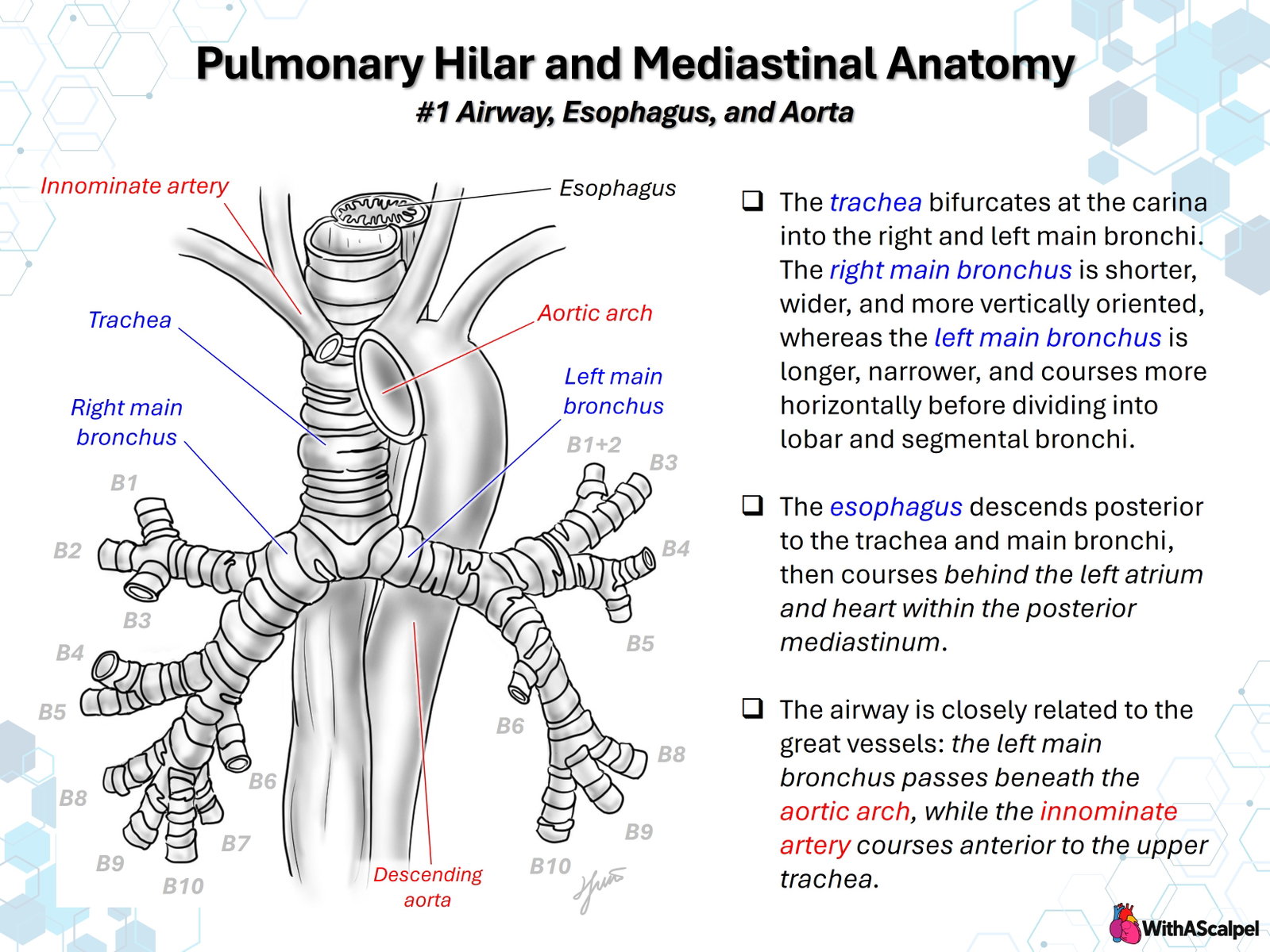
The pulmonary hila and mediastinum form a compact three-dimensional interface between the conducting airways, esophagus, heart, and great vessels. Their relationships are best understood in three planes: the tracheobronchial tree extends inferiorly and laterally; the esophagus descends posterior to the central airway; and the aortic arch curves over the left main bronchus before continuing as the descending thoracic aorta. The brachiocephalic (innominate) artery crosses the anterior aspect of the upper intrathoracic trachea in the usual left-sided aortic arch configuration.
These relationships explain normal imaging appearances and determine where congenital vascular anomalies, postoperative changes, or enlarged cardiovascular structures may compress the airway or esophagus. Anatomical descriptions refer to the usual configuration; the actual course, caliber, and level of contact vary with age, thoracic geometry, and congenital anatomy.
2. Trachea, Carina, and Main Bronchi
The trachea is supported anteriorly and laterally by C-shaped cartilaginous rings and posteriorly by a membranous wall containing the trachealis muscle. Its distal bifurcation, the carina, separates the right and left main bronchi. In adults, this junction is typically near the sternal angle and T4–T5 level, although its position changes with respiration and development. A cadaveric microanatomical study showed that carinal cartilage may incorporate the distal tracheal ring and contributions from either main bronchus, emphasizing that the carina is not a uniformly constructed isolated cartilage [1].
The right main bronchus is generally shorter, wider, and more vertically aligned with the trachea. The right upper lobe bronchus arises relatively proximally; the remaining bronchus continues as the bronchus intermedius before supplying the middle and lower lobes. A deeply advanced endotracheal tube can preferentially enter the right main bronchus and may obstruct the early right upper lobe orifice. This familiar adult asymmetry is less pronounced or more variable in some infants and young children, so tube position must be assessed rather than inferred from bronchial angles alone.
The left main bronchus is longer and courses more horizontally, passing beneath the aortic arch toward the left hilum. It lies anterior to the esophagus and near the descending aorta, while the left pulmonary artery crosses superior and anterior to the bronchial pathway at the hilum. The left main bronchus divides into upper- and lower-lobe bronchi without a right-sided equivalent of the bronchus intermedius.
3. Bronchopulmonary Segmental Organization
Each lobar bronchus divides into segmental bronchi that supply anatomically recognizable bronchopulmonary segments. The classic segmental framework remains useful for bronchoscopy, cross-sectional imaging, surgical localization, and interpreting the distribution of atelectasis or infection [2].
On the right, the upper lobe contains B1 (apical), B2 (posterior), and B3 (anterior); the middle lobe contains B4 (lateral) and B5 (medial); and the lower lobe is conventionally described by B6 (superior) and basal branches B7–B10. On the left, the upper lobe commonly has a combined B1+2 (apicoposterior) branch and B3 (anterior), with B4 and B5 forming the lingular bronchi. The left lower lobe has B6 and basal branches, often without a discrete B7.
These labels describe common patterns, not invariant anatomy. Bronchoscopic series document considerable variation in segmental origins and branching, including accessory, absent, fused, and displaced branches [3]. When an intervention depends on segment identification, the actual bronchial tree on CT or bronchoscopy takes precedence over a schematic B1–B10 arrangement.
4. Esophageal Course and Relationship to the Left Atrium
The esophagus begins posterior to the larynx and trachea and enters the thorax in the posterior mediastinal compartment. Its cervical and superior thoracic course is generally behind the airway, although it may deviate laterally. At the level of the tracheal bifurcation, it is posterior to the main bronchi and adjacent mediastinal connective tissues. More inferiorly, it descends behind the left atrium, separated from the cardiac chamber by pericardial and variable intervening soft tissue layers, before traversing the diaphragmatic hiatus.
The close association of the posterior left atrial wall and esophagus is particularly important. CT studies in adult populations demonstrate that the esophageal course relative to the posterior left atrium and pulmonary venous ostia is variable, with broad areas of close apposition [4,5]. Consequently, a single schematic relationship should not be treated as a patient-specific map. These adult imaging data establish the principle of anatomical variability but should not be extrapolated quantitatively to infants.
Clinically, the posterior position of the esophagus explains why extrinsic cardiovascular compression may produce dysphagia, feeding intolerance, or an impression on contrast esophagography. The same proximity is relevant to transesophageal echocardiography, esophageal instrumentation, and procedures involving the posterior left atrium. In complex congenital anatomy, the course of the esophagus should be interpreted in relation to the entire vascular configuration rather than to the left atrium alone.
5. Aortic Arch, Brachiocephalic Artery, and Descending Aorta
In the usual left aortic arch, the ascending aorta continues superiorly and posteriorly into the transverse arch, which passes over the left main bronchus and then turns inferiorly as the descending thoracic aorta. The descending aorta initially lies to the left of the vertebral column and is closely related to the posterior left hilar region and esophagus. The left main bronchus therefore occupies a constrained space beneath the arch, anterior to deeper posterior mediastinal structures.
The brachiocephalic artery is ordinarily the first branch of the arch. It travels obliquely upward toward the right and crosses anterior to the trachea. Its relationship to the airway depends on the height and course of the vessel, tracheal position, and surrounding support. An unusually low or posterior crossing can cause focal anterior tracheal compression, particularly in young children with compliant airways. Innominate artery compression represents a vascular-related airway compression syndrome, not a complete vascular ring [6].
The ligamentum arteriosum typically connects the proximal left pulmonary artery region to the undersurface of the arch near the isthmus. The left recurrent laryngeal nerve loops beneath the arch near this ligamentous complex, making it an important structure during arch or vascular ring dissection. In right-sided arch variants, the ligamentum and aberrant subclavian artery may complete a ring around both the trachea and esophagus. These are spatial relationships with direct consequences for operative exposure, safe mobilization, and residual compression.
6. Three-Dimensional Relationships at the Hila
The term hilum describes the point of entry and exit of the bronchus, pulmonary artery, pulmonary veins, bronchial vessels, nerves, and lymphatics. The pulmonary root is the connective-tissue-covered bundle that traverses the mediastinal pleura. The spatial arrangement is different on the two sides and cannot be represented adequately by a single anterior view.
On the right, the upper lobe bronchus commonly originates superior to the pulmonary artery at the hilum—the eparterial bronchus—whereas the bronchus intermedius and subsequent branches are hyparterial. On the left, the pulmonary artery generally lies superior to the left main bronchus and its lobar branches, which are hyparterial. Pulmonary veins occupy relatively anterior and inferior positions within the root, although their individual branching and drainage patterns may vary.
The arch lies superior to the left main bronchus; the left pulmonary artery is more anterior at the hilum; and the descending aorta and esophagus lie in the posterior mediastinal plane. This combination helps orient axial CT images and explains why abnormalities of different vascular structures produce compression at different levels. For example, an enlarged pulmonary artery may deform a bronchus from an anterior or superior direction, whereas an arch anomaly may narrow it from a superior or posterior direction. The precise vector must be verified on patient imaging.
7. Congenital Vascular Relationships and Compression
The standard arrangement becomes particularly clinically relevant when the aortic arch or pulmonary arteries develop abnormally. Double aortic arch can surround the trachea and esophagus, creating circumferential constraint. A right aortic arch with aberrant left subclavian artery and left ligamentum arteriosum may form another complete ring. The retroesophageal vessel or associated Kommerell diverticulum may contribute to posterior esophageal or airway compression. Imaging reviews demonstrate that these configurations cannot be reliably characterized by arch sidedness alone [7,8].
A left pulmonary artery sling has a different mechanism: the anomalous left pulmonary artery typically arises from the right pulmonary artery and passes between the trachea and esophagus to reach the left lung. This may compress the distal trachea or right main bronchus and can coexist with intrinsic tracheal stenosis and complete cartilage rings. The sling is not the same as a conventional aortic vascular ring.
An important distinction is external compression versus intrinsic airway weakness. Chronic extrinsic pressure can coexist with tracheomalacia or bronchomalacia; relieving the vascular constraint does not necessarily restore normal dynamic airway stability immediately. The diagnostic and operative questions are therefore separate: which structure compresses the airway, and how much intrinsic or persistent dynamic obstruction remains? Multidisciplinary evaluation may be necessary when symptoms and static imaging do not agree [6,8].
8. Imaging and Endoscopic Assessment
Echocardiography is valuable for associated intracardiac disease, arch sidedness, and selected vascular connections, but may not completely depict the airway–esophagus–vascular interface. CT angiography with multiplanar and three-dimensional reconstruction is particularly useful for mapping arch branches, the descending aorta, pulmonary arteries, and focal airway narrowing within one dataset. Images should be inspected in axial, sagittal, and coronal planes; a visually compelling 3D rendering must not replace careful assessment of the source images [9,10].
For suspected aerodigestive compression, a systematic imaging review should identify the carina; the narrowest tracheal or bronchial segment; the length and direction of narrowing; the adjacent compressing vessel; the course of the esophagus; and any associated arch, pulmonary arterial, or tracheal anomaly. An esophagram may help define functional indentation or passage disturbance. MRI angiography provides an alternative vascular assessment without ionizing radiation, although the choice depends on age, motion, acquisition needs, and local expertise [6,7].
Dynamic flexible bronchoscopy evaluates airway shape, pulsatile indentation, secretions, and expiratory collapse, providing physiological information that static CT may miss. Sedation level, airway pressure, and respiratory phase influence dynamic findings. Bronchoscopy and angiographic imaging are complementary: bronchoscopy characterizes luminal behavior, whereas cross-sectional imaging defines the external structures responsible for its deformation [6].
9. Implications for Procedures and Postoperative Care
Accurate airway–vascular orientation matters during intubation, bronchoscopy, aortic arch reconstruction, vascular ring repair, pulmonary artery mobilization, and esophageal instrumentation. Left main bronchial compression should be considered when airway narrowing occurs beneath the arch or adjacent to an enlarged left pulmonary artery. Unilateral ventilation or unexpected lobar atelectasis may result from endotracheal tube malposition, a displaced segmental orifice, external compression, or dynamic bronchomalacia; these mechanisms require different corrective actions.
Persistent wheezing, stridor, ventilator dependence, or extubation failure after successful cardiovascular reconstruction should not automatically be attributed to residual vascular obstruction. Intrinsic tracheobronchomalacia and fixed stenosis may remain clinically important. Published pediatric cardiac surgical experience associates tracheobronchomalacia with complex postoperative respiratory management, supporting targeted airway assessment when the clinical course is disproportionate to the cardiac findings [11].
10. Key Clinical Principles
- Think in three dimensions: the esophagus is posterior to the central airway, while the aortic arch passes over the left main bronchus.
- Respect bronchial asymmetry: the right main bronchus is usually more vertical, but pediatric angles and segmental origins are variable.
- Identify the anterior crossing vessel: the brachiocephalic artery may produce localized tracheal compression without forming a complete vascular ring.
- Do not confuse mechanisms: aortic vascular rings, pulmonary artery sling, intrinsic stenosis, and airway malacia are distinct, although they may coexist.
- Use complementary modalities: CT or MRI delineates external anatomy; dynamic bronchoscopy assesses airway behavior.
- Interpret persistent symptoms anatomically: residual compression, fixed narrowing, and dynamic collapse require different management strategies.
References
- Kim CY, Rizzuto K, Tahan D, et al. Anatomy and variations of the carina and its cartilaginous makeup: a cadaveric study. Surgical and Radiologic Anatomy. 2025. DOI: 10.1007/s00276-025-03579-y. PMID: 39904909.
- Boyden EA. A synthesis of the prevailing patterns of the bronchopulmonary segments in the light of their variations. Diseases of the Chest. 1949. DOI: 10.1378/CHEST.15.6.657. PMID: 18139478.
- Beder S, Küpeli E, Karnak D, Kayacan O. Tracheobronchial variations in Turkish population. Clinical Anatomy. 2008;21:531–538. DOI: 10.1002/ca.20667. PMID: 18698650.
- Lemola K, Sneider MB, Desjardins B, et al. Computed tomographic analysis of the anatomy of the left atrium and the esophagus: implications for left atrial catheter ablation. Circulation. 2004. DOI: 10.1161/01.CIR.0000149714.31471.FD. PMID: 15569839.
- Tsao HM, Wu MH, Higa S, et al. Anatomic relationship of the esophagus and left atrium: implication for catheter ablation of atrial fibrillation. Chest. 2005. DOI: 10.1378/CHEST.128.4.2581. PMID: 16236927.
- Chiu P, Zendejas B, Baird C. Multidisciplinary approach to vascular rings and vascular-related aerodigestive compression: a clinical practice review. Translational Pediatrics. 2023. DOI: 10.21037/tp-23-39. PMID: 37427058.
- Dillman JR, Attili AK, Agarwal PP, et al. Common and uncommon vascular rings and slings: a multi-modality review. Pediatric Radiology. 2011. DOI: 10.1007/s00247-011-2131-2. PMID: 21594540.
- Backer CL, Monge MC, Popescu AR, et al. Vascular rings. Seminars in Pediatric Surgery. 2016. DOI: 10.1053/j.sempedsurg.2016.02.009. PMID: 27301603.
- Lee EY, Siegel MJ. MDCT of tracheobronchial narrowing in pediatric patients. Journal of Thoracic Imaging. 2007. DOI: 10.1097/RTI.0b013e3180cab6e8. PMID: 17721348.
- Lambert V, Sigal-Cinqualbre A, Belli E, et al. Preoperative and postoperative evaluation of airways compression in pediatric patients with 3-dimensional multislice computed tomographic scanning: effect on surgical management. Journal of Thoracic and Cardiovascular Surgery. 2005. DOI: 10.1016/J.JTCVS.2004.08.030. PMID: 15867788.
- Chen Q, Langton-Hewer S, Marriage S, et al. Influence of tracheobronchomalacia on outcome of surgery in children with congenital heart disease and its management. Annals of Thoracic Surgery. 2009. DOI: 10.1016/j.athoracsur.2009.08.039. PMID: 19932270.