1. Overview
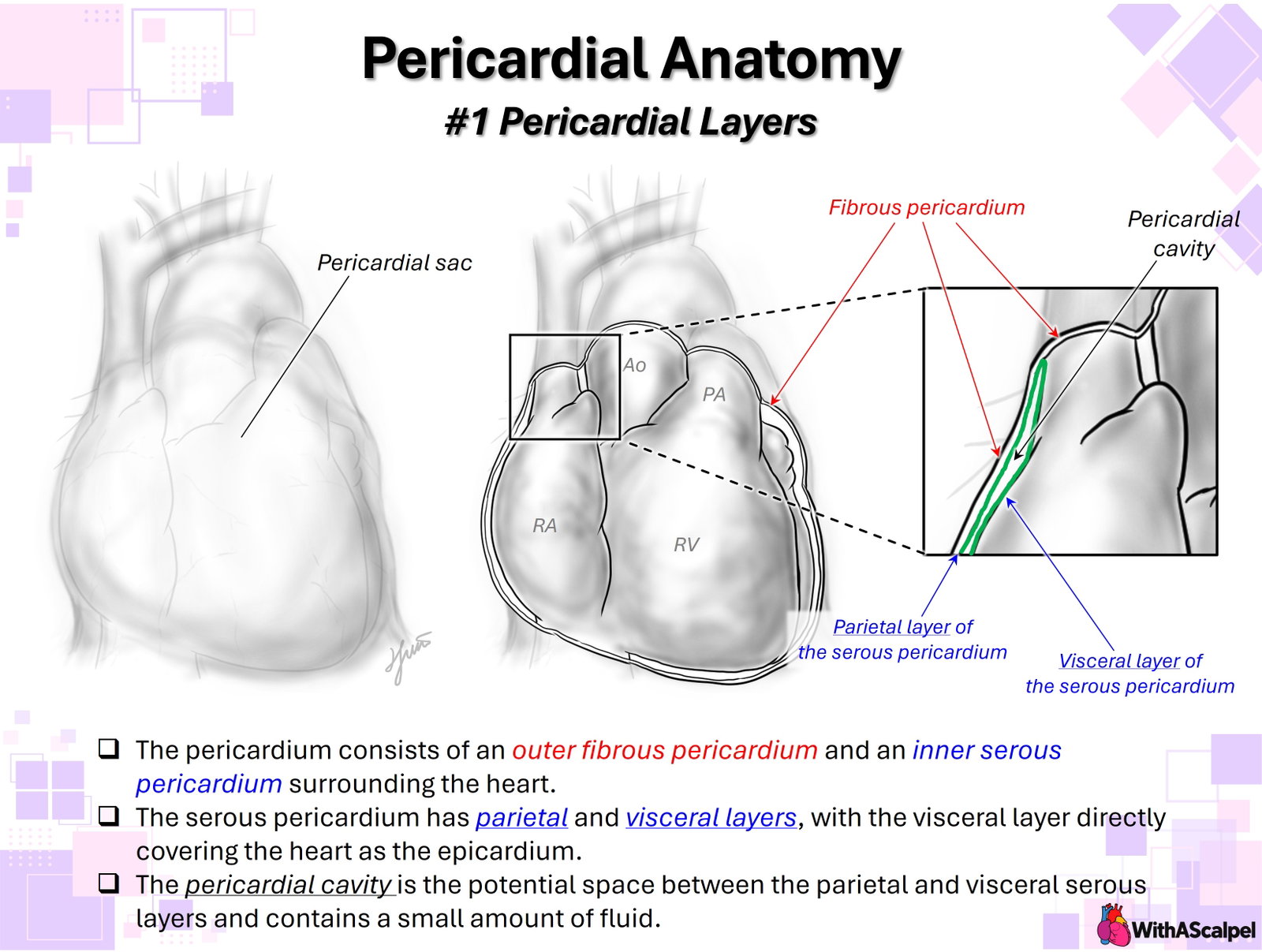
The pericardium is a layered fibroserous envelope surrounding the heart and the intrapericardial portions of the great vessels. Its organization is conceptually simple but clinically and surgically important: an outer fibrous pericardium forms the mechanically robust supporting sac, whereas an inner serous pericardium provides a smooth mesothelial surface. The serous component is continuous but is conventionally divided into a parietal layer, which lines the inner surface of the fibrous sac, and a visceral layer, which directly covers the heart and is termed the epicardium. The narrow potential space between these two serous surfaces is the pericardial cavity. [1,2]
Figure 1. Pericardial layers. The outer fibrous pericardium encloses the serous pericardium; the parietal and visceral serous layers face the pericardial cavity, and the visceral layer directly covers the heart as the epicardium.
This arrangement explains several features that are otherwise easy to confuse. The fibrous and parietal serous layers effectively move together as the outer wall of the pericardial sac, whereas the visceral serous layer moves with the heart. At the roots of the great vessels, the serous membrane reflects from the cardiac surface onto the parietal surface, creating the pericardial reflections, sinuses, and recesses. These reflections are not merely descriptive anatomy; they define important planes around the ascending aorta, pulmonary trunk, caval veins, pulmonary veins, and left atrium. [1,3]
A useful three-dimensional model is to imagine the serous pericardium as a closed membrane that has been invaginated by the developing heart and great vessels. The heart therefore lies within the fibrous pericardial sac but, strictly speaking, outside the serous cavity itself: only the opposing mesothelial surfaces face the cavity. This relationship is analogous to that between the lung and pleural cavity.
The arterial reflection surrounds the ascending aorta and pulmonary trunk as they emerge from the heart. The venous reflections surround the superior and inferior caval veins and pulmonary veins. Because the arterial and venous reflections are separated, a passage persists between them—the transverse pericardial sinus. The oblique sinus forms as a cul-de-sac within the venous reflection behind the left atrium. [1,6]
In operative terminology, “pericardium” often refers primarily to the fibrous sac together with its adherent parietal serosa, whereas “epicardium” refers to the visceral serosa on the cardiac surface. This practical terminology is appropriate provided the continuity of the serous membrane is understood.
2. Fibrous Pericardium
The fibrous pericardium is the mechanically robust outer component of the sac. Histologically, it consists predominantly of collagen with elastic fibers arranged in interlacing bundles, producing substantial tensile strength while retaining limited deformability. Human histologic studies demonstrate a collagen-rich fibrous component beneath the serosal surface and an external connective-tissue layer that blends with surrounding mediastinal attachments. [1,4]
Inferiorly, the fibrous pericardium is firmly related to the central tendon of the diaphragm. Superiorly, it becomes continuous with the adventitial connective tissue surrounding the great vessels. Anterior connections to the sternum and surrounding mediastinal connective tissues contribute to stabilization of the heart within the middle mediastinum. These attachments do not immobilize the heart; rather, they limit excessive displacement while accommodating normal cardiac motion and physiologic changes in volume. [1,2]
Laterally, the fibrous pericardium is closely related to the mediastinal pleura. The phrenic nerves, accompanied by the pericardiacophrenic vessels, descend along its lateral surfaces anterior to the pulmonary hila. This relationship is particularly important during cardiac and thoracic surgery because lateral pericardial traction, cautery, or an improperly positioned incision can injure the phrenic nerve. Cadaveric study confirms the intimate relationship between the thoracic phrenic nerve and its pericardiacophrenic vascular supply. [5]
3. Serous Pericardium
3.1 Parietal Layer
The parietal layer of the serous pericardium is closely adherent to the inner surface of the fibrous pericardium. Although commonly illustrated as a distinct sheet, the two layers are not readily separable during normal gross dissection. The parietal serosa presents a smooth mesothelial surface toward the pericardial cavity, permitting the beating heart to move against the surrounding sac with minimal friction. [1,4]
Microscopically, the luminal surface consists of mesothelial cells overlying a thin submesothelial connective-tissue layer. Ultrastructural examination of normal human parietal pericardium demonstrates mesothelial microvilli, intercellular junctions, and an underlying collagen-rich fibrous framework. These features support both mechanical durability and transport of fluid across the serosal membrane. [4]
3.2 Visceral Layer: The Epicardium
The visceral layer of the serous pericardium is the epicardium, forming the outer surface of the heart. It consists of mesothelium and underlying connective tissue whose thickness varies regionally. Epicardial adipose tissue, coronary arteries and veins, autonomic nerves, and lymphatic structures occupy the subepicardial compartment, particularly within the atrioventricular and interventricular grooves. Thus, the epicardium is not simply a friction-reducing surface; it overlies the pathways of major coronary and neural structures. [1,2]
The visceral and parietal layers are continuous at the pericardial reflections around the great vessels. Consequently, the “two layers” of the serous pericardium should not be conceptualized as independent membranes with free margins. They are different regions of one continuous mesothelial surface.
4. Pericardial Reflections, Sinuses, and Recesses
The transition from visceral to parietal serous pericardium occurs around the arterial and venous roots. These reflections generate spaces whose configuration reflects the relationships among the great arteries, atria, systemic veins, and pulmonary veins. Anatomical and imaging studies demonstrate appreciable normal variability in their geometry. [6–8]
The transverse pericardial sinus lies posterior to the ascending aorta and pulmonary trunk and anterior to the atrial roof and adjacent venous structures. Surgically, it provides a natural passage behind the great arterial trunks and can facilitate mobilization or encirclement of the ascending aorta and pulmonary artery. Its extensions form several recesses around the great vessels. [6,7]
This anatomy is also important radiologically. Fluid extending into the transverse sinus or one of its recesses can simulate a mediastinal cyst, lymph node, or other soft-tissue lesion on cross-sectional imaging if the expected pericardial reflections are not recognized. [6,7]
The oblique pericardial sinus is a blind recess posterior to the left atrium. It is formed primarily by reflections surrounding the pulmonary veins and inferior vena cava and communicates inferiorly with the general pericardial cavity. Contemporary CT analysis demonstrates substantial interindividual variation in the size, depth, entrance, and configuration of both the transverse and oblique sinuses. The classic textbook configuration therefore serves as an anatomical framework rather than an invariant geometry. [8]
5. Pericardial Cavity and Pericardial Fluid
The pericardial cavity is the potential space between the parietal and visceral serous layers. Under normal conditions, only a small volume of serous fluid—on the order of several tens of milliliters in adults—is distributed as a thin film between the mesothelial surfaces. [2,3,9]
Pericardial fluid is derived predominantly from plasma ultrafiltration across the serosal microvasculature and is cleared mainly through lymphatic pathways. Mesothelial cells also participate in fluid transport and local homeostasis. The fluid and mesothelial surfaces together provide lubrication, allowing repeated cardiac movement with minimal friction between the epicardium and parietal serosa. [3,9]
The pericardial cavity should therefore be considered a dynamic serosal interface, not simply an empty compartment. When fluid accumulates, it distributes within the main cavity and along the pericardial recesses according to gravity, patient position, adhesions, and local anatomy. The hemodynamic consequence of an effusion depends not only on its absolute volume but also on its rate of accumulation and the pressure-volume characteristics of the fibrous sac.
6. Developmental Basis of the Epicardium
The epicardium has a developmental origin distinct from the myocardium. It arises predominantly from the proepicardial organ, an extracardiac mesothelial structure associated with the venous pole of the embryonic heart. Proepicardial cells reach and spread across the myocardial surface, eventually establishing the epicardial mesothelium. [10]
A subset of epicardial cells undergoes epithelial-to-mesenchymal transition and contributes to several cellular populations within the developing heart, particularly cardiac fibroblasts and vascular smooth-muscle lineages. Epicardial-derived cells and epicardial signaling also participate in coronary vascular development and myocardial maturation. [10]
These developmental functions should be distinguished from mature gross anatomical terminology. In the postnatal heart, visceral serous pericardium and epicardium refer to the same anatomical surface layer, despite the broader developmental and biological roles of epicardial-derived cells.
7. Functional Significance of the Layered Arrangement
The pericardium performs several interacting functions. First, its mesothelial surfaces and pericardial fluid minimize friction. Second, the fibrous sac provides mechanical protection and contributes to stabilization of the heart and proximal great vessels. Third, because the fibrous pericardium has limited acute distensibility, it restrains sudden increases in total cardiac volume and contributes to ventricular interdependence. [2,11]
At normal filling volumes, pericardial restraint is modest. As cardiac volume increases, however, the pressure-volume relationship of the pericardial sac becomes progressively steeper. Expansion of one chamber may then influence filling of another because both ventricles occupy the same constrained intrapericardial space. Experimental and physiologic studies form the basis for this concept and help explain the marked importance of pericardial constraint in acute chamber dilation, cardiac tamponade, and constrictive physiology. [11]
The pericardium is therefore neither essential for immediate survival nor physiologically irrelevant. Its hemodynamic influence is relatively subtle in the normal euvolemic state but becomes increasingly important when cardiac volume or pericardial contents change rapidly.
8. Surgical and Imaging Relevance
For the surgeon, the fundamental distinction is between the fibrous/parietal pericardial sac and the visceral epicardial surface. After median sternotomy, opening the fibrous pericardium enters the pericardial cavity; traction sutures placed in the fibrous edges can create a pericardial well and improve exposure. In lateral approaches, the course of the phrenic nerve on the fibrous pericardium should be identified before the pericardiotomy is extended. [5]
The serous reflections determine how far the great vessels can be mobilized intrapericardially and where dissection transitions into extrapericardial tissue. The transverse sinus provides a surgically useful plane behind the great arterial trunks, whereas the oblique sinus identifies the posterior pericardial space behind the left atrium.
Reoperations fundamentally alter this anatomy. Inflammation and postoperative healing can replace the normally smooth, fluid-lubricated interface with adhesions between the epicardium and parietal pericardium. Structures that were previously separated by a potential space may therefore become inseparable scarred planes, accounting for much of the difficulty and risk of redo cardiac exposure. Mesothelial injury and repair are central to this process. [3]
Cross-sectional imaging similarly depends on understanding normal pericardial anatomy. CT and CMR can delineate pericardial thickness, calcification, fluid, fat, reflections, and recesses, whereas echocardiography is particularly valuable for determining the hemodynamic consequences of effusion and pericardial constraint. Recognition of normal recesses prevents physiologic pericardial fluid from being mistaken for pathological mediastinal structures. [6,7]
Key Clinical Principles
- Fibrous outside, serous inside. The fibrous pericardium provides the mechanically strong supporting sac; the serous pericardium provides its mesothelial surfaces.
- Parietal and visceral serosa are continuous. Their reflections around the great vessels create the transverse and oblique sinuses and associated recesses.
- The visceral serous layer is the epicardium. It covers the heart and overlies subepicardial coronary vessels, adipose tissue, nerves, and lymphatics.
- The pericardial cavity is a potential space. A thin film of fluid normally separates its serosal surfaces and permits low-friction cardiac motion.
- The phrenic nerve is a critical lateral surgical landmark. It courses along the fibrous pericardium with the pericardiacophrenic vessels.
- Pericardial anatomy is functional anatomy. The same structure that permits cardiac mobility also restrains acute expansion and contributes to ventricular interaction.
- Pericardial reflections matter in surgery and imaging. The sinuses and recesses should be understood as three-dimensional spaces created by serosal folding rather than as simple lines on a diagram.
References
- Rodriguez ER, Tan CD. Structure and anatomy of the human pericardium. Prog Cardiovasc Dis. 2017;59(4):327–340. doi:10.1016/j.pcad.2016.12.010. PMID: 28062264. (PubMed)
- Hoit BD. Anatomy and physiology of the pericardium. Cardiol Clin. 2017;35(4):481–490. doi:10.1016/j.ccl.2017.07.002. PMID: 29025540. (PubMed)
- Jaworska-Wilczynska M, Trzaskoma P, Szczepankiewicz AA, Hryniewiecki T. Pericardium: structure and function in health and disease. Folia Histochem Cytobiol. 2016;54(3):121–125. doi:10.5603/FHC.a2016.0014. PMID: 27654013. (PubMed)
- Ishihara T, Ferrans VJ, Jones M, Boyce SW, Kawanami O, Roberts WC. Histologic and ultrastructural features of normal human parietal pericardium. Am J Cardiol. 1980;46(5):744–753. doi:10.1016/0002-9149(80)90424-5. PMID: 7435384. (PubMed)
- Jiang S, Xu WD, Shen YD, Xu JG, Gu YD. An anatomical study of the full-length phrenic nerve and its blood supply: clinical implications for endoscopic dissection. Anat Sci Int. 2011;86(4):225–231. doi:10.1007/s12565-011-0114-x. PMID: 21993978. (PubMed)
- Choe YH, Im JG, Park JH, Han MC, Kim CW. The anatomy of the pericardial space: a study in cadavers and patients. AJR Am J Roentgenol. 1987;149(4):693–697. doi:10.2214/AJR.149.4.693. PMID: 3498317. (PubMed)
- Żurada A, Ustymowicz A, Loukas M, Michalak M, Czyżewska D, Gielecki J. Computerized tomography of the transverse pericardial sinus: normal or pathologic? Clin Anat. 2017;30(1):61–70. doi:10.1002/ca.22778. PMID: 27578603. (PubMed)
- Burysz M, Batko J, Olejek W, et al. Morphology and anatomical classification of pericardial cavities: oblique and transverse sinuses. J Clin Med. 2023;12(13):4320. doi:10.3390/jcm12134320. PMID: 37445356. (PubMed)
- Vogiatzidis K, Zarogiannis SG, Aidonidis I, et al. Physiology of pericardial fluid production and drainage. Front Physiol. 2015;6:62. doi:10.3389/fphys.2015.00062. PMID: 25852564. (PubMed)
- Männer J, Pérez-Pomares JM, Macías D, Muñoz-Chápuli R. The origin, formation and developmental significance of the epicardium: a review. Cells Tissues Organs. 2001;169(2):89–103. doi:10.1159/000047867. PMID: 11399849. (PubMed)
- Watkins MW, LeWinter MM. Physiologic role of the normal pericardium. Annu Rev Med. 1993;44:171–180. doi:10.1146/annurev.me.44.020193.001131. PMID: 8476238. (PubMed)