1. Introduction
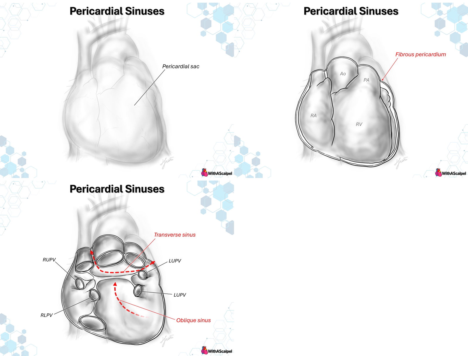
The pericardial sinuses are extensions of the pericardial cavity created where the serous pericardium reflects around vessels entering and leaving the heart. They are not separate chambers or defects in the pericardial sac. Rather, they are continuous potential spaces whose configuration follows the arrangement of the arterial and venous attachments of the heart.
Two sinuses have particular anatomical and clinical importance. The transverse pericardial sinus courses behind the ascending aorta and pulmonary trunk, separating the arterial and venous poles. The oblique pericardial sinus occupies a blind recess behind the left atrium, bounded by reflections around the pulmonary veins and inferior vena cava (IVC). Understanding the difference is essential for interpreting imaging, navigating surgical planes, and recognizing fluid collections in unusual locations [1,2].
2. The Pericardial Sac and Serous Reflections
2.1 Fibrous and serous layers
The fibrous pericardium is the outer connective-tissue envelope. It is anchored inferiorly to the central tendon of the diaphragm and blends superiorly with connective tissue surrounding the great vessels. The parietal serous pericardium lines its inner surface; the visceral serous pericardium, or epicardium, invests the heart. The intervening cavity normally contains a thin film of lubricating fluid [1].
The sinuses are formed by reflections of the serous layers, not by folds of the fibrous pericardium itself. The fibrous sac provides an outer boundary; the reflections define where the parietal surface becomes continuous with the epicardial covering of the vessels and heart.
2.2 Arterial and venous attachments
The arterial reflection surrounds the proximal ascending aorta and pulmonary trunk. The venous reflection is more complex, following the superior vena cava (SVC), IVC, and pulmonary venous entrances. The space left between these two systems forms the transverse sinus. Within the venous reflection, the posterior pericardial cavity extends upward behind the left atrium as the oblique sinus [2].
A useful three-dimensional concept is that the heart is attached to the pericardium at an arterial hilum and a venous hilum. Intact-pericardium dissections demonstrate that the geometry of these attachments is more complex than a simple pair of circular openings. The extent of epicardial coverage over the atrial roof, in particular, varies with the configuration of the reflections [2].
3. Transverse Pericardial Sinus
3.1 Boundaries and course
The transverse sinus is a passage from one side of the pericardial cavity to the other, located posterior to the ascending aorta and pulmonary trunk. The venous structures and superior atrial surfaces form its posterior relationships. The SVC is an important landmark near the rightward entrance, while the roof of the left atrium and the right pulmonary artery are important posterior and superior neighbors, respectively [2,5].
The rightward opening is approached between the ascending aorta and SVC. Toward the left, the passage emerges adjacent to the pulmonary trunk and left atrial appendage. It is important to distinguish structures forming the boundary of a sinus from structures actually lying within its lumen: the right pulmonary artery, for example, is a close anatomical neighbor, not a free structure traversing the pericardial space.
3.2 Recesses and variable morphology
The transverse sinus has irregular contours and extensions around the arterial roots, including superior and inferior aortic recesses and pulmonary-related recesses. Consequently, a cross-sectional image may show several apparently disconnected fluid pockets even though they belong to one communicating pericardial system.
A CT study of 121 adults classified transverse-sinus morphology as concave, wine-type, straight, or convex, with the concave configuration most common. These are descriptive morphological categories, not validated predictors of surgical difficulty or disease [4]. Their practical lesson is that the sinus should not be assumed to form an equally wide, straight tunnel in every patient.
3.3 Operative significance
The transverse sinus provides a recognizable route behind the arterial trunks and can assist in encircling or controlling the great arteries. However, a tape or instrument should be passed only after the surgeon identifies the correct vessel and confirms that the posterior plane is free. Congenital malposition of the great arteries, prior surgical adhesions, and distorted mediastinal anatomy may substantially alter this relationship.
During pulmonary artery banding, for example, the transverse sinus may facilitate passage behind the main pulmonary artery. The surgeon must still confirm the plane, avoid the pulmonary arterial wall and adjacent structures, and position the band away from the pulmonary valve and branch pulmonary artery origins. Anatomical familiarity does not justify blind instrumentation.
4. Oblique Pericardial Sinus
4.1 A blind recess behind the left atrium
Unlike the transverse sinus, the oblique sinus is a cul-de-sac. It opens inferiorly into the general pericardial cavity and extends superiorly behind the left atrium, ending at the superior venous reflection. Its anterior wall is the posterior epicardial surface of the left atrium, and its posterior boundary is the parietal pericardium overlying the posterior mediastinal structures [1–3].
The right and left pulmonary venous reflections form its lateral boundaries. The IVC reflection contributes to the inferior-right aspect of the venous attachment. Consequently, the space is defined primarily by the pulmonary venous entrances and their surrounding serous reflections, rather than by the external outline of the left atrium.
4.2 Relationships to the pulmonary veins
The right upper and right lower pulmonary veins enter the left atrium on its right posterior aspect; the corresponding left upper and left lower veins lie along the left side. Their pericardial sleeves and intervening recesses vary in length and configuration. In 107 cadaveric dissections, Chaffanjon and colleagues documented variations in pulmonary venous segments and venous pericardial reflections, supporting a range of posterior pericardial morphologies [3].
The oblique sinus is therefore not a uniformly deep cavity. In the 121-patient CT study, oblique sinuses were classified as shallow or deep, each with a narrow or wide entrance; the shallow, narrow-entrance type was most common [4]. These observations arise primarily from adult imaging and anatomy and should not be extrapolated as dimensional standards for children.
4.3 Clinical and operative relevance
Introducing a hand or instrument superiorly behind the heart from the inferior pericardial cavity enters the oblique sinus and brings it against the posterior left atrial surface. This location is relevant to posterior atrial exposure, pulmonary venous mobilization, and selected approaches to anomalous pulmonary venous connection.
The posterior pericardium also lies close to the esophagus, vagal branches, and autonomic neural structures. Anatomical studies emphasize that the extent of pericardial coverage and the position of these structures vary [2,5]. Accordingly, dissection beyond the venous reflections requires deliberate identification of the operative plane; the apparent posterior extent of the sinus is not a guarantee of a safe, avascular corridor.
5. Other Pericardial Recesses
Smaller recesses arise as the serous membrane follows individual vessels. Relevant examples include the superior aortic recess, the postcaval recess adjacent to the SVC, the left pulmonic recess near the left pulmonary artery and left upper pulmonary vein, and recesses between the pulmonary veins. These are normal extensions of the pericardial cavity and may become conspicuous when filled with fluid [6].
The distinction between the transverse and oblique sinuses remains fundamental: the transverse sinus provides a superior communicating passage behind the arterial trunks, whereas the oblique sinus terminates superiorly behind the left atrium. They do not communicate directly through the intervening venous pericardial reflection, although both open into the broader pericardial cavity [2].
6. Imaging and Diagnostic Interpretation
6.1 Echocardiography
Echocardiography is the initial investigation for most suspected pericardial effusions. Fluid within the transverse sinus may appear posterior to the ascending aorta or pulmonary trunk, while fluid in the oblique sinus can appear immediately behind the left atrium. Epicardial fat, neighboring mediastinal structures, or localized collections may mimic a mass on an isolated plane. Serial views and attention to anatomical continuity help distinguish these findings [8].
Transesophageal echocardiography may improve visualization of the posterior left atrium and adjacent pericardial space when transthoracic windows are limited. Nevertheless, a single echocardiographic image cannot always define the full extent of a complex recess or postoperative compartment.
6.2 Computed tomography and magnetic resonance imaging
CT provides a detailed map of the relationships between the pericardial recesses, great vessels, pulmonary veins, and mediastinal lymph nodes. In a retrospective study of 941 CT examinations, Kodama and colleagues found that thin sections depicted both large and small recesses more frequently than thicker sections, underscoring the influence of acquisition technique on apparent anatomy [7].
A fluid-filled recess may be mistaken for adenopathy, a cystic lesion, a vascular abnormality, or—in selected locations—an aortic dissection. Helpful discriminators include fluid attenuation, the expected anatomical location, continuity with the pericardial cavity on multiplanar imaging, and lack of an enhancing solid component [6,9].
Cardiac magnetic resonance can complement CT by assessing fluid characteristics, pericardial inflammation, and cardiac functional consequences. Contemporary imaging guidance supports modality selection according to the clinical question, rather than routine use of every technique [10]. The sinus itself is normal anatomy; fluid in a recess is not automatically pathological.
7. Implications for Cardiac Surgery and Interventions
7.1 Exposure and vessel control
Safe use of the transverse sinus begins with visual identification of the ascending aorta, pulmonary trunk, SVC, and the expected posterior plane. Gentle blunt development may permit passage of a clamp, dissector, or tape. Resistance should prompt reassessment rather than forceful advancement. In a reoperative chest, normal pericardial planes may be obliterated by adhesions.
7.2 Posterior atrial and pulmonary venous procedures
The oblique sinus helps orient the surgeon to the posterior left atrium. For pulmonary venous reconstruction, the relevant distinction is between mobilizing the surrounding pericardial reflection and dissecting directly against the venous wall. Excessive traction or circumferential dissection can compromise venous geometry, particularly where a short pulmonary venous segment enters the atrium.
Epicardial and thoracoscopic ablation procedures also use the transverse and oblique sinuses to position guide catheters and energy-delivery devices around the posterior left atrium. Published technical experience describes access through both spaces; these procedures additionally require attention to the phrenic nerves, coronary vessels, esophagus, and adjacent atrial tissue [5,11]. Such techniques should not be generalized to altered congenital anatomy without individual assessment.
8. Postoperative Collections and Regional Tamponade
Surgery, bleeding, inflammatory exudate, and adhesions can convert a freely communicating pericardial cavity into partially isolated compartments. Fluid or hematoma may accumulate adjacent to either sinus or behind the atria. The resulting pressure can cause regional cardiac compression without the classic appearance of a circumferential effusion [12].
A CT series of 33 patients with postoperative pericardial effusion identified localized compartmental distributions in 16 cases. This historical, selected cohort illustrates the anatomical mechanism, not the prevalence of regional tamponade in contemporary cardiac surgery [12]. Case literature also documents hemodynamically important localized collections that were difficult to recognize on transthoracic echocardiography but evident on CT [13].
When postoperative low cardiac output, unexplained elevation of filling pressures, or chamber compression raises concern, imaging should actively examine the posterior pericardial space as well as the anterior and circumferential regions. Drainage decisions depend on the collection's location, consistency, accessibility, and hemodynamic effects. A drain positioned in one compartment may not decompress another.
9. Key Anatomical and Clinical Principles
- Serous reflections create the sinuses; the fibrous pericardium forms the surrounding sac.
- Transverse sinus: a communicating passage posterior to the ascending aorta and pulmonary trunk.
- Oblique sinus: a blind superiorly directed recess posterior to the left atrium, bounded by venous reflections.
- Pulmonary venous sleeves are variable; preserve venous geometry during mobilization.
- Fluid in a normal recess can mimic pathology; use multiplanar anatomical relationships to resolve uncertainty.
- Postoperative adhesions alter communication; a localized collection can produce regional tamponade.
- Surgical navigation must be anatomy-specific; do not assume that the normal sinus plane remains patent.
References
- Rodriguez ER, Tan CD. Structure and anatomy of the human pericardium. Prog Cardiovasc Dis. 2017;59:327–340. DOI: 10.1016/j.pcad.2016.12.010. PMID: 28062264.
- Mori S, Hanna P, Dacey MJ, et al. Comprehensive anatomy of the pericardial space and the cardiac hilum: Anatomical dissections with intact pericardium. JACC Cardiovasc Imaging. 2022;15:927–942. DOI: 10.1016/j.jcmg.2021.04.016. PMID: 34147441.
- Chaffanjon P, Brichon PY, Faure C, Favre JJ. Pericardial reflection around the venous aspect of the heart. Surg Radiol Anat. 1997;19:17–21. DOI: 10.1007/BF01627729. PMID: 9060112.
- Burysz M, Batko J, Olejek W, et al. Morphology and anatomical classification of pericardial cavities: Oblique and transverse sinuses. J Clin Med. 2023;12:4320. DOI: 10.3390/jcm12134320. PMID: 37445356.
- Lachman N, Syed FF, Habib A, et al. Correlative anatomy for the electrophysiologist, Part I: The pericardial space, oblique sinus, transverse sinus. J Cardiovasc Electrophysiol. 2010;21:1421–1426. DOI: 10.1111/j.1540-8167.2010.01872.x. PMID: 20731740.
- Levy-Ravetch M, Auh YH, Rubenstein WA, et al. CT of the pericardial recesses. AJR Am J Roentgenol. 1985;144:707–714. DOI: 10.2214/ajr.144.4.707. PMID: 3872026.
- Kodama F, Fultz PJ, Wandtke JC. Comparing thin-section and thick-section CT of pericardial sinuses and recesses. AJR Am J Roentgenol. 2003;181:1101–1108. DOI: 10.2214/ajr.181.4.1811101. PMID: 14500240.
- Penmasta S, Silbiger JJ. The transverse and oblique sinuses of the pericardium: Anatomic and echocardiographic insights. Echocardiography. 2019;36:170–176. DOI: 10.1111/echo.14211. PMID: 30506589.
- Ferreira HDP, Erasmus LT, Strange TA, et al. Pericardial recesses on computed tomography: Implications for the pulmonologist. Clin Chest Med. 2024;45:237–248. DOI: 10.1016/j.ccm.2024.02.002. PMID: 38816085.
- Klein AL, Wang TKM, Cremer PC, et al. Pericardial diseases: International position statement on new concepts and advances in multimodality cardiac imaging. JACC Cardiovasc Imaging. 2024;17:937–988. DOI: 10.1016/j.jcmg.2024.04.010. PMID: 39111992.
- Verberkmoes N, Olsthoorn J, Dekker L. Minimally invasive totally thoracoscopic stand-alone surgical ablation for atrial fibrillation. Multimed Man Cardiothorac Surg. 2020. DOI: 10.1510/mmcts.2020.011. PMID: 32436664.
- Duvernoy O, Larsson SG, Persson K, et al. Pericardial effusion and pericardial compartments after open heart surgery: An analysis by computed tomography and echocardiography. Acta Radiol. 1990;31:41–46. PMID: 2340224.
- Vohra HA, Khout H, Bapu D, et al. Localised pericardial tamponade diagnosed by computed tomography: A case presentation. J Med Case Rep. 2007;1:162. DOI: 10.1186/1752-1947-1-162. PMID: 18053151.