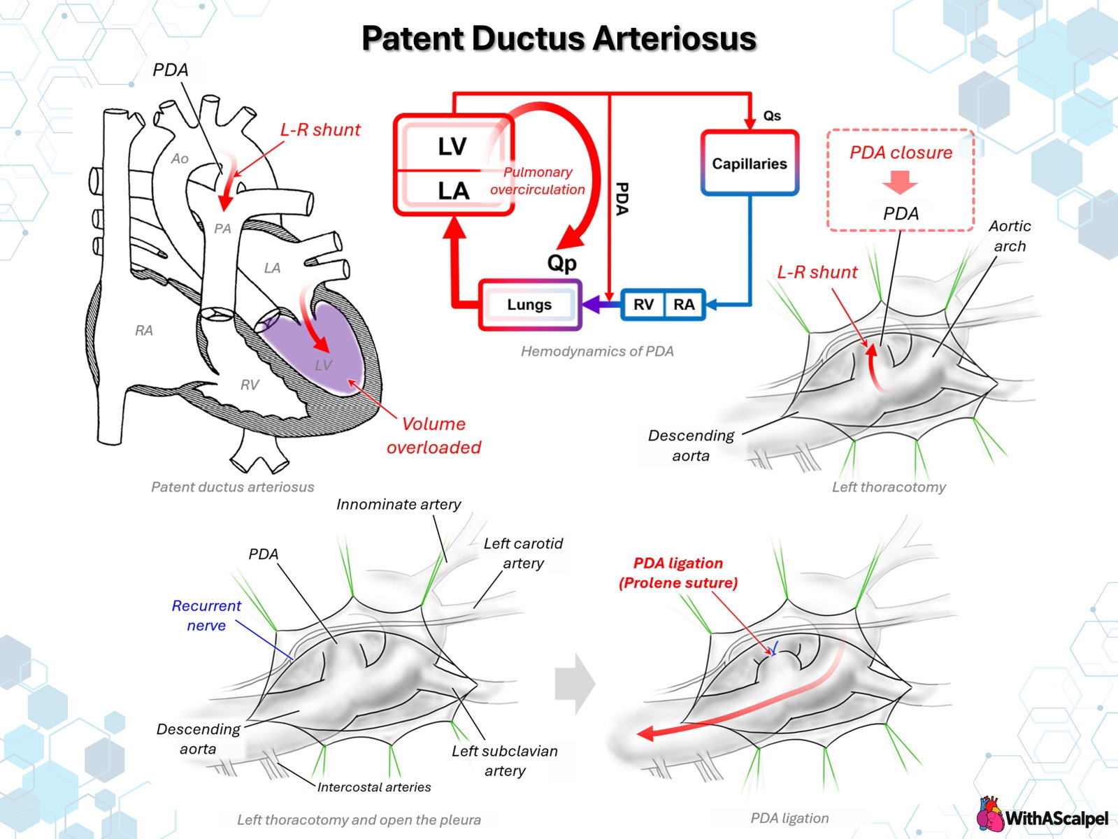
Patent ductus arteriosus (PDA) is persistence after birth of the fetal vascular connection between the pulmonary artery and the proximal descending aorta. Its clinical importance is determined less by the mere presence of ductal patency than by the magnitude and direction of shunting, pulmonary vascular resistance, systemic vascular resistance, and the capacity of the myocardium to accommodate the resulting volume load. A hemodynamically important PDA produces pulmonary overcirculation and increased pulmonary venous return, creating predominantly left atrial and left ventricular volume overload.[1,2]
Management spans observation, pharmacologic treatment in selected preterm infants, transcatheter occlusion, and surgical closure. In contemporary practice, transcatheter closure is preferred for most anatomically suitable infants and children and is increasingly feasible even in extremely low-birth-weight infants. Surgical ligation remains an important definitive option when catheter closure is unsuitable, unsuccessful, unavailable, or judged to carry unacceptable anatomical risk.[2-5]
1. Ductal Anatomy and Postnatal Transition
The ductus arteriosus is derived from the distal portion of the left sixth aortic arch. It connects the pulmonary arterial circulation—typically near the junction of the main and left pulmonary arteries—to the proximal descending thoracic aorta just distal to the left subclavian artery.[1]
During fetal life, pulmonary vascular resistance (PVR) is high and pulmonary blood flow is relatively low. The ductus therefore carries predominantly right-to-left flow from the pulmonary artery into the descending aorta, allowing a substantial portion of right ventricular output to bypass the unexpanded lungs.
After birth, lung expansion and oxygenation cause a rapid decline in PVR. Simultaneously, removal of the placental circulation increases systemic vascular resistance. Increased arterial oxygen tension, decreased circulating prostaglandin concentrations, and intrinsic ductal smooth-muscle contraction normally produce functional ductal constriction followed by anatomical remodeling and permanent closure.[1,2]
Persistence of an open ductus after these transitional changes exposes the pulmonary artery to blood flowing from the higher-pressure systemic circulation.
2. Hemodynamics of the PDA
2.1 Development of the left-to-right shunt
Once systemic pressure exceeds pulmonary pressure, flow through an unrestricted PDA becomes predominantly aorta-to-pulmonary artery, or left-to-right. Because aortic pressure exceeds pulmonary arterial pressure during both systole and diastole in a patient without severe pulmonary hypertension, ductal flow is typically continuous.[1]
The magnitude of the shunt is determined by several interacting variables:
- ductal diameter, length, and geometry;
- systemic-to-pulmonary pressure gradient;
- PVR relative to systemic vascular resistance;
- ventricular function;
- associated congenital cardiac lesions.
A small restrictive PDA may transmit a high-velocity jet but generate little additional pulmonary blood flow. Conversely, a large nonrestrictive PDA allows pulmonary flow to be determined largely by the relative downstream vascular resistances.
2.2 Pulmonary overcirculation
Blood entering the pulmonary artery through the PDA adds directly to effective right ventricular pulmonary output. Consequently,
Qp = effective systemic venous pulmonary flow + ductal left-to-right shunt.
The additional pulmonary flow traverses the pulmonary capillary bed and returns through the pulmonary veins to the left atrium. The hemodynamic burden therefore appears principally as increased LA and LV preload, rather than primary right ventricular volume overload.[1,2]
As the ductal shunt becomes larger, echocardiography typically demonstrates progressive LA and LV dilation. Pulmonary venous and left atrial pressures may rise, particularly when ventricular compliance or systolic reserve is limited, contributing to pulmonary edema and respiratory symptoms.
2.3 Systemic runoff
The PDA is also a pathway for systemic arterial blood to escape into the lower-resistance pulmonary circulation, especially during diastole. A large shunt can therefore produce a low diastolic arterial pressure and widened pulse pressure.
This concept becomes particularly important in premature neonates, in whom substantial ductal runoff may compromise perfusion of systemic vascular beds. Retrograde or reduced diastolic flow can be demonstrated in the descending aorta and, in severe cases, in mesenteric, renal, or cerebral circulations.[3]
Thus, a large PDA should not be conceptualized simply as “too much pulmonary flow.” It creates a coupled physiology of pulmonary overcirculation plus potentially reduced effective systemic perfusion.
3. Clinical Consequences
The spectrum ranges from an incidentally detected small PDA to severe cardiopulmonary compromise.
A moderate or large PDA may produce tachypnea, feeding intolerance, poor weight gain, recurrent respiratory symptoms, or clinical heart failure. Examination may reveal a continuous infraclavicular murmur, hyperdynamic precordium, bounding peripheral pulses, and widened pulse pressure. Very large PDAs with elevated pulmonary artery pressure may have a less characteristic murmur because the instantaneous aortic-to-pulmonary pressure gradient is reduced.[1,2]
Chronic exposure to excessive pulmonary blood flow and pressure can promote pulmonary vascular remodeling. With advanced pulmonary vascular disease, the left-to-right shunt decreases, becomes bidirectional, and may ultimately reverse. Once irreversible pulmonary vascular disease with right-to-left ductal shunting is established, routine closure is inappropriate because the ductus may function as an important pressure-relief pathway for the right ventricle.
Accordingly, large PDAs associated with significant pulmonary hypertension require careful assessment of pulmonary vascular resistance and shunt direction rather than closure based simply on ductal size.
4. Echocardiographic Assessment
Echocardiography is the principal diagnostic modality in infants and children. Assessment should extend beyond confirmation of ductal patency.
Important findings include:
- ductal size and morphology;
- direction and velocity profile of ductal flow;
- LA and LV dimensions;
- evidence of pulmonary overcirculation;
- pulmonary artery pressure estimates;
- descending aortic diastolic flow reversal;
- ventricular systolic function;
- associated congenital lesions.
A large LA or LV provides physiological evidence that the ductus is producing substantial recirculation through the pulmonary vascular bed. Conversely, the absence of left-heart dilation in an otherwise uncomplicated term infant or child generally argues against a major chronic left-to-right shunt.
In premature infants, defining a “hemodynamically significant PDA” is more complex. Ductal diameter alone is insufficient; contemporary neonatal assessment integrates ductal characteristics with cardiac loading, systemic perfusion, respiratory status, and the overall clinical trajectory.[3]
5. Indications and Methods for Closure
5.1 Transcatheter closure
Transcatheter occlusion has largely replaced surgery for anatomically suitable PDAs in infants, children, and adults.[2] Advances in low-profile delivery systems have extended catheter-based closure to very small premature infants.
In the prospective multicenter Amplatzer Piccolo trial, 200 patients weighing at least 700 g were enrolled, including 100 weighing 2 kg or less. Implant success was 95.5% overall and 99% in those ≤2 kg, with high rates of complete closure.[4] Subsequent three-year follow-up supported sustained ductal occlusion and a low incidence of serious device-related complications.[5]
Important device-related concerns include protrusion into the descending aorta or left pulmonary artery, vascular injury, device embolization, tricuspid-valve injury during catheter manipulation, and residual shunting. These risks are particularly relevant in extremely small infants and depend heavily on anatomy and operator experience.
5.2 Role of surgical closure
Surgical closure remains relevant when:
- catheter anatomy is unfavorable;
- device placement is unsuccessful;
- vascular access or patient size creates prohibitive catheter risk;
- urgent definitive closure is required and catheter therapy is unavailable;
- another thoracic operation provides an opportunity for simultaneous treatment.
Surgical PDA closure can usually be performed without cardiopulmonary bypass through a left thoracotomy. Thoracoscopic clipping is another established approach in selected patients, although institutional experience varies.[6-8]
6. Surgical Anatomy
The operation is fundamentally an exercise in identifying the ductus while protecting the structures surrounding the distal aortic arch.
Through the left chest, the principal landmarks are the:
- distal aortic arch;
- descending thoracic aorta;
- left subclavian artery;
- pulmonary artery;
- PDA;
- left recurrent laryngeal nerve.
The left recurrent laryngeal nerve is particularly important. After branching from the vagus nerve, it courses beneath the aortic arch adjacent to the ductal region before ascending in the tracheoesophageal groove. Excessive traction, thermal injury, clip entrapment, or aggressive circumferential ductal dissection can produce vocal-cord paresis or paralysis.
The ductus itself may be extremely thin-walled and fragile, particularly in premature infants. Dissection must therefore balance adequate visualization with avoidance of unnecessary manipulation.
7. Surgical PDA Ligation
7.1 Exposure
The patient is positioned in the right lateral decubitus position. A limited left posterolateral or lateral thoracotomy is performed, usually through an upper intercostal space appropriate to the patient's size.
After entering the pleural cavity, the lung is gently displaced anteriorly. The descending aorta is identified and followed superiorly toward the distal arch. The left subclavian artery provides another useful landmark.
The mediastinal pleura overlying the ductal region is opened carefully. Exposure should permit clear definition of the descending aorta, arch, and PDA before occlusion is attempted.
7.2 Identification and protection of the recurrent laryngeal nerve
The relationship between the recurrent nerve and the ductus should be actively considered rather than assumed. The nerve may be difficult to visualize directly in very small infants.
Dissection immediately around the superior and posterior ductal surfaces should therefore be conservative. Electrocautery near the nerve should be minimized, and traction on tissues surrounding the ductus should be gentle.
7.3 Ductal occlusion
Several surgical methods are used, including nonabsorbable suture ligation and clip application. The precise method depends on patient size, ductal morphology, tissue quality, and surgeon preference.
When suture ligation is used, adequate visualization must be obtained to ensure that the suture encompasses the ductus rather than an adjacent structure. Secure complete occlusion is required, but excessive tension on a fragile neonatal ductus should be avoided.
Thoracoscopic approaches generally achieve interruption using clips and can reduce the size of the thoracic incision. Published series demonstrate high closure rates, although conversion, residual shunting, recurrent laryngeal nerve injury, and chylothorax remain recognized complications.[6-8]
8. Completion Assessment and Complications
Immediately after ligation, the surgeon should assess for:
- absence of significant hemorrhage;
- complete ductal occlusion;
- intact aortic and pulmonary arterial flow;
- preservation of surrounding structures.
Postoperative echocardiography confirms the absence of residual ductal flow and evaluates ventricular function.
Important complications include:
- left recurrent laryngeal nerve injury;
- residual PDA;
- ductal or aortic hemorrhage;
- pneumothorax;
- chylothorax;
- wound complications;
- postoperative hemodynamic deterioration.
Recurrent laryngeal nerve dysfunction is particularly relevant in premature infants, in whom even unilateral vocal-cord dysfunction may affect extubation, airway protection, and feeding.
9. Hemodynamics After Closure
PDA closure produces an abrupt change in ventricular loading conditions. Before closure, the LV ejects partly into the systemic circulation and partly through the low-resistance pulmonary runoff pathway. Closure eliminates that pathway.
Two changes therefore occur simultaneously:
- LV preload falls because pulmonary overcirculation and pulmonary venous return decrease.
- Effective LV afterload rises because all LV output must now enter the systemic circulation.
In most children this transition is well tolerated. In premature infants with a large PDA and limited myocardial reserve, however, acute LV systolic dysfunction, systemic hypotension, increased ventilatory requirements, and low cardiac output may develop—a constellation often termed post-ligation cardiac syndrome.[9]
Recent comparative observational data suggest that this syndrome occurs more frequently after surgical ligation than after transcatheter closure in very-low-weight infants, although patient selection and baseline illness severity complicate direct causal comparisons.[9,10]
Key Clinical and Surgical Principles
- A PDA is fundamentally a recirculation lesion. Aortic blood returns to the pulmonary circulation and then to the LA and LV.
- The principal chronic cardiac volume load is left-sided. Significant shunting produces LA/LV dilation through increased pulmonary venous return.
- Shunt magnitude depends on resistance as well as anatomy. Falling PVR after birth converts ductal patency into progressively important left-to-right flow.
- Large PDAs can compromise systemic perfusion. Diastolic runoff is especially important in premature neonates.
- Transcatheter closure is now the dominant definitive therapy when anatomy permits.
- Surgery remains a reliable non-CPB option when catheter therapy is unsuitable or unsuccessful.
- The left recurrent laryngeal nerve is the key neural structure at risk during surgical ligation.
- Closure abruptly changes LV loading conditions. Post-closure ventricular dysfunction and hypotension should be anticipated, particularly in premature infants with large shunts.
Figure. Patent ductus arteriosus: pathophysiology and surgical closure. After postnatal reduction in pulmonary vascular resistance, a PDA permits left-to-right flow from the aorta to the pulmonary artery, increasing pulmonary blood flow and pulmonary venous return and producing LA/LV volume overload. Surgical closure through a left thoracic approach exposes the ductus between the pulmonary artery and descending aorta. The relationship of the left recurrent laryngeal nerve to the ductal region is critical during dissection and ligation.
References
- Schneider DJ, Moore JW. Patent ductus arteriosus. Circulation. 2006. DOI: 10.1161/CIRCULATIONAHA.105.592063. PMID: 17060397.
- Schneider DJ. The patent ductus arteriosus in term infants, children, and adults. Semin Perinatol. 2012. DOI: 10.1053/j.semperi.2011.09.025. PMID: 22414886.
- Hamrick SEG, Sallmon H, Rose AT, et al. Patent ductus arteriosus of the preterm infant. Pediatrics. 2020. DOI: 10.1542/peds.2020-1209. PMID: 33093140.
- Sathanandam SK, Gutfinger D, O'Brien L, et al. Amplatzer Piccolo Occluder clinical trial for percutaneous closure of the patent ductus arteriosus in patients ≥700 grams. Catheter Cardiovasc Interv. 2020. DOI: 10.1002/ccd.28973. PMID: 32433821.
- Morray BH, Sathanandam S, Forbes T, et al. 3-year follow-up of a prospective, multicenter study of the Amplatzer Piccolo Occluder for transcatheter patent ductus arteriosus closure in children ≥700 grams. J Perinatol. 2023. DOI: 10.1038/s41372-023-01741-1. PMID: 37587183.
- Vida VL, Rubino M, Bottio T, et al. Thoracoscopic closure of the patent arterial duct. Cardiol Young. 2004. DOI: 10.1017/S1047951104002082. PMID: 15691405.
- Vanamo K, Berg E, Kokki H, Tikanoja T. Video-assisted thoracoscopic versus open surgery for persistent ductus arteriosus. J Pediatr Surg. 2006. DOI: 10.1016/j.jpedsurg.2006.03.002. PMID: 16818053.
- Stankowski T, Aboul-Hassan S, Marczak J, Szymańska A, Augustyn C, Cichoń R. Minimally invasive thoracoscopic closure versus thoracotomy in children with patent ductus arteriosus. J Surg Res. 2017. DOI: 10.1016/j.jss.2016.08.097. PMID: 27993195.
- Duboue P, Padovani P, Bouteiller X, et al. Post-ligation cardiac syndrome after surgical versus transcatheter closure of patent ductus arteriosus in low body weight premature infants: a multicenter retrospective cohort study. Eur J Pediatr. 2024. DOI: 10.1007/s00431-024-05481-y. PMID: 38381375.
- Tabb C, Aggarwal S, Bajaj M, Natarajan G. Comparative effectiveness of surgical ligation and catheter closure of patent ductus arteriosus in preterm infants. Pediatr Cardiol. 2023. DOI: 10.1007/s00246-023-03199-6. PMID: 37316609.