Fontan Circulation: Integrated Physiology, Complications, and Rescue Strategies
1. Introduction
The Fontan circulation is the definitive stage of surgical palliation for patients with a functionally single ventricle when biventricular repair is not feasible. Its defining feature is total cavopulmonary connection without a subpulmonary ventricle: systemic venous blood passes through the pulmonary arteries and lungs before returning to the pulmonary venous atrium and the single systemic ventricle. By separating systemic and pulmonary venous streams, Fontan completion generally improves systemic arterial oxygenation and eliminates much of the ventricular volume load associated with earlier palliation. These benefits are achieved at the cost of chronically elevated systemic venous pressure and limited ventricular preload reserve [1,2].
The essential physiologic trade-off is therefore not simply correction of cyanosis. Fontan circulation converts a parallel, often volume-loaded circulation into a circulation arranged largely in series, in which the pulmonary vascular bed becomes the principal obstacle to systemic venous return. Long-term function depends on the interaction of pulmonary vascular impedance, unobstructed venous pathways, ventricular filling properties, atrioventricular (AV) valve competence, and lymphatic drainage.
2. Transition From First-Stage Palliation to Glenn and Fontan Circulation
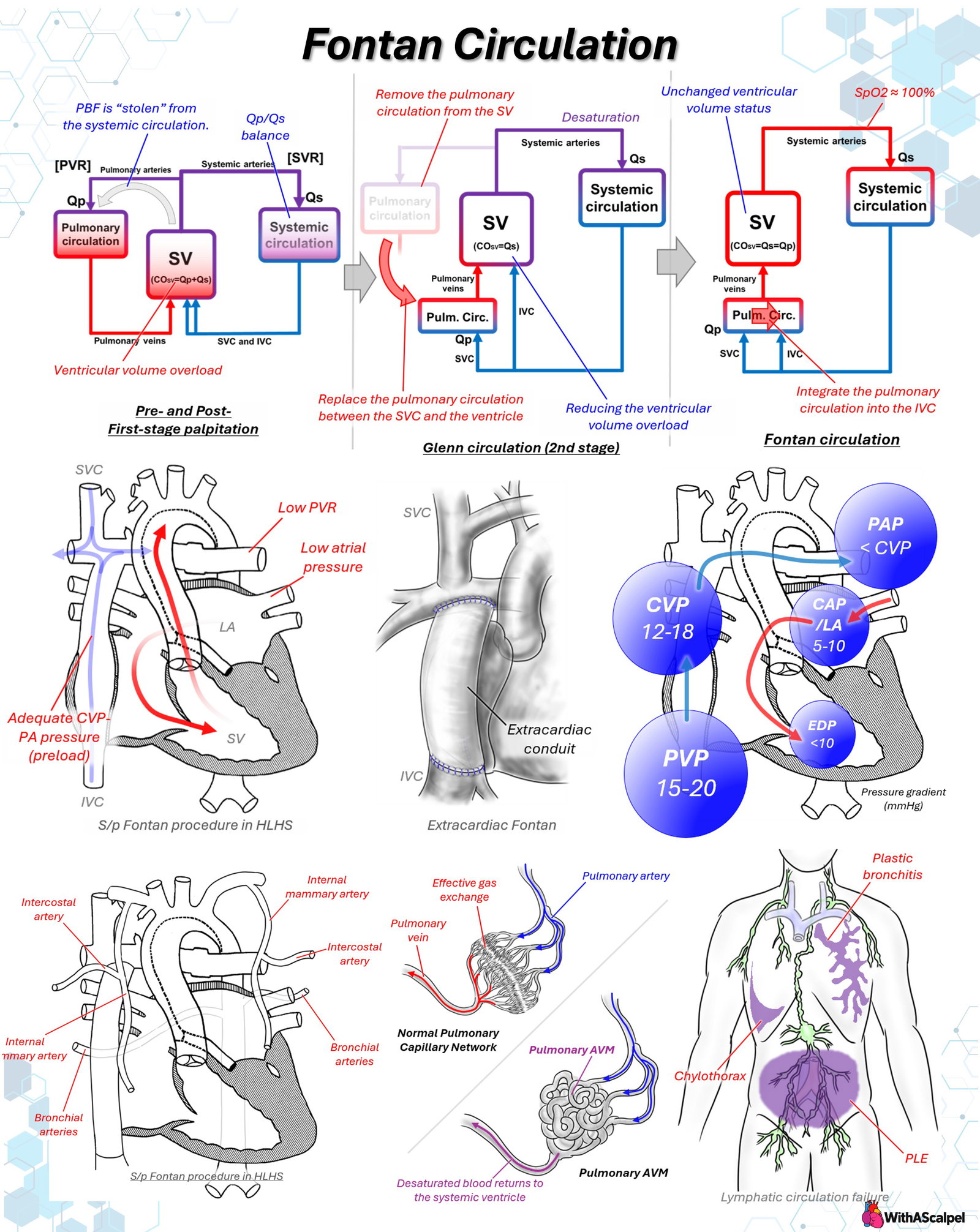
First-stage circulation. Following neonatal palliation, pulmonary blood flow may arise from a systemic-to-pulmonary shunt, a right ventricle–to–pulmonary artery conduit, a patent ductus arteriosus, or native antegrade pulmonary outflow. When the single ventricle supplies both systemic and pulmonary circulations, total ventricular output approximately equals systemic flow plus pulmonary flow. Excessive pulmonary blood flow competes with effective systemic perfusion and increases pulmonary venous return and ventricular volume loading. Consequently, an apparently satisfactory systemic oxygen saturation can coexist with inadequate systemic oxygen delivery.
Bidirectional Glenn circulation. Superior vena caval (SVC) blood is connected directly to the pulmonary arteries, while inferior vena caval (IVC) return continues through the heart. The Glenn reduces the fraction of systemic venous blood returning directly to the ventricle and substantially decreases ventricular volume loading. However, the IVC contribution to systemic venous return generally remains unsaturated, so systemic arterial desaturation persists. The Glenn circulation also provides an opportunity to evaluate pulmonary vascular development, ventricular function, and AV valve competence before Fontan completion [1].
Fontan completion. An extracardiac conduit or intracardiac lateral tunnel incorporates IVC and hepatic venous return into the pulmonary circulation. In an extracardiac Fontan, a conduit connects the divided IVC to the pulmonary artery confluence; a lateral tunnel redirects IVC flow through an intra-atrial baffle. Both configurations establish total cavopulmonary connection. The reconstruction must be widely patent, avoid conduit compression or angulation, and distribute hepatic venous effluent to both lungs when possible. Conduit diameter and the decision to create a fenestration depend on patient size, anatomy, expected flow, and institutional strategy.
3. Pressure Gradients and the Fontan Hemodynamic Bottleneck
In the absence of a right-sided pumping chamber, systemic venous pressure supplies the driving pressure for pulmonary blood flow. The relationship may be approximated as:
Fontan pressure − pulmonary venous atrial pressure ≈ pulmonary blood flow × total cavopulmonary and pulmonary vascular resistance.
This equation emphasizes that the measured pressure difference incorporates both intrinsic pulmonary vascular resistance (PVR) and energy losses from the Fontan pathway, pulmonary artery geometry, and pulmonary venous connections. The systemic ventricle powers the systemic arterial circulation; it does not independently generate pressure proximal to the pulmonary arteries. Ventricular diastolic pressure remains important because it determines the downstream filling pressure against which the Fontan pathway must operate [2].
A clinically stable Fontan commonly exhibits a resting central venous pressure in the low-to-mid teens, with some patients approaching 18 mmHg or more. Pulmonary artery pressure is similar to, or slightly lower than, upstream Fontan pressure unless there is pathway obstruction. Pulmonary venous atrial pressure and ventricular end-diastolic pressure are ideally low. The source diagrams illustrate venous pressures around 12–18 mmHg, atrial pressures around 5–10 mmHg, and transpulmonary pressure differences of several millimeters of mercury. These figures are illustrative, not universal reference ranges or standalone intervention thresholds.
Elevated venous pressure with low atrial pressure suggests excessive resistance within the cavopulmonary pathway or pulmonary vascular bed. When both venous and atrial pressures are elevated, ventricular diastolic dysfunction, systolic dysfunction, AV valve regurgitation, pulmonary venous obstruction, or multiple lesions must be considered. A discrete obstruction can be clinically important even when its measured gradient is small because the Fontan circuit operates with limited pressure and energy reserves.
Preload deprivation limits stroke volume during exercise, dehydration, vasodilation, tachyarrhythmia, and positive-pressure ventilation. Conversely, indiscriminate fluid administration may increase systemic venous congestion without materially improving ventricular filling. Negative intrathoracic pressure during spontaneous inspiration generally assists pulmonary venous transit, whereas excessive positive intrathoracic pressure may impede flow. Ventilatory management nevertheless requires balancing this physiology against lung recruitment and the work of breathing.
4. Patient Selection and High-Risk Hemodynamics
Pre-Fontan assessment should establish unobstructed cavopulmonary and pulmonary venous pathways, favorable pulmonary artery dimensions, low pulmonary vascular impedance, acceptable ventricular filling pressure, preserved systemic ventricular function, and no important AV valve regurgitation. Echocardiography, cardiovascular magnetic resonance, and catheterization provide complementary information; catheterization is particularly informative when direct pressure measurements, PVR estimation, or treatment of residual lesions is required.
Published cohorts identify elevated pulmonary artery pressure, impaired ventricular function, and unfavorable anatomy as markers of failure to complete Fontan palliation [3,4]. In a 2026 multicenter study, pre-Fontan mean pulmonary artery pressure was associated continuously with subsequent death or transplantation, without a clear single threshold separating low and high risk [3]. Therefore, proposed cutoffs such as indexed PVR above 3 Wood units·m² or ventricular end-diastolic pressure above 12 mmHg should be interpreted in context, not treated as absolute contraindications. Prior pulmonary artery or pulmonary venous interventions, significant AV valve disease, syndromic conditions, respiratory support requirements, and poor functional status warrant integrated assessment.
5. Fenestration: A Deliberate Decompression Pathway
A Fontan fenestration provides a controlled right-to-left communication from the conduit or lateral tunnel to the atrium. When pulmonary vascular impedance or ventricular filling pressure becomes limiting, blood can bypass the pulmonary bed, augment systemic ventricular preload, and partially decompress the Fontan pathway. The trade-off is reduced arterial saturation and exposure to paradoxical embolization.
Fenestration changes the balance between oxygen saturation and cardiac output. A patient with modest desaturation but substantially improved systemic flow may achieve better systemic oxygen delivery than one with higher saturation and severe low-output physiology. The usefulness of this mechanism depends on the amount of shunting and the patient's pulmonary vascular and ventricular conditions; fenestration cannot correct a major mechanical obstruction.
Evidence does not establish routine fenestration as superior for every patient. A meta-analysis involving 19 studies and 4,806 patients reported less need for prolonged pleural drainage with fenestration but no significant reduction in overall Fontan failure [5]. A smaller randomized trial of 40 extracardiac Fontan patients did not demonstrate a clear reduction in pleural effusion burden [6]. These findings support selective, anatomy- and risk-based use rather than a universal approach. If later closure is considered, temporary test occlusion should assess cardiac output, Fontan pressure, and ventricular filling before permanent device closure.
6. Systemic-to-Pulmonary Collaterals and Ventricular Volume Loading
Aortopulmonary collateral arteries commonly arise from intercostal, internal thoracic, thyrocervical, and bronchial arterial territories. They deliver systemic arterial blood to the pulmonary vascular bed independently of cavopulmonary venous flow. This additional pulmonary flow returns through the pulmonary veins and increases systemic ventricular preload and volume work.
The collateral circuit may be hemodynamically inefficient: systemic ventricular output is recirculated through the lungs rather than being delivered to systemic tissue beds. Extensive collateral flow can contribute to elevated atrial filling pressure, pleural effusions, and persistent ventricular dilation. Phase-contrast cardiac MRI can estimate collateral flow from the difference between pulmonary venous return and flow through the branch pulmonary arteries [7].
Catheter embolization is considered when substantial collateral burden contributes to adverse physiology, but the effect of routine prophylactic occlusion on long-term outcomes remains uncertain. Collaterals are not interchangeable with pulmonary arteriovenous malformations or venovenous collaterals; the source, direction, and destination of the shunt determine its hemodynamic consequences.
7. Pulmonary Arteriovenous Malformations and Hepatic Flow Distribution
Pulmonary arteriovenous malformations (PAVMs) create intrapulmonary right-to-left shunting, allowing blood to traverse abnormal vascular channels without effective alveolar gas exchange. The resulting desaturation can resemble that associated with Fontan fenestration, but PAVMs do not provide direct decompression of the Fontan venous pathway.
PAVMs are particularly associated with exclusion of hepatic venous blood from the pulmonary arteries after superior cavopulmonary connection or Kawashima-type circulation. Clinical observations support an important protective role for hepatic venous effluent, although the responsible circulating factor or combination of factors has not been definitively identified [8]. PAVMs can persist or arise after Fontan completion when hepatic venous flow is preferentially directed to only one lung, particularly in complex heterotaxy anatomy [9].
Evaluation should distinguish PAVMs from fenestration, systemic venovenous collaterals, residual intracardiac shunting, and pulmonary parenchymal disease. Depending on the mechanism, management may require reconstruction or redirection of hepatic venous flow rather than simple occlusion of individual diffuse lesions.
8. Lymphatic Dysfunction: A Consequence of Venous Hypertension
Systemic venous hypertension impedes lymphatic drainage into the central veins while increasing hepatic and intestinal lymph production. Abnormal lymphatic anatomy and retrograde collateral pathways may redirect lymph toward the pleural cavity, bronchial tree, or intestinal mucosa. Elevated pressure alone does not explain every manifestation; lymphatic flow distribution and abnormal connections are critical determinants [10,11].
The principal clinical phenotypes are chylothorax, characterized by lymphatic accumulation in the pleural space; plastic bronchitis, characterized by bronchial casts related to abnormal lymphatic leakage; and protein-losing enteropathy, characterized by intestinal protein loss, hypoalbuminemia, edema, and impaired nutritional or immune status.
Assessment begins with Fontan hemodynamics, ventricular function, AV valve competence, pathway patency, and possible thrombosis. T2-weighted MR lymphatic imaging and dynamic contrast MR lymphangiography can identify lymphatic congestion and abnormal flow pathways. Management may combine reduction of venous pressure, treatment of a correctable obstruction, nutritional and medical therapy, and anatomy-directed lymphatic embolization or decompression. Such interventions are specialized and are supported primarily by observational experience rather than randomized trials [10,11].
9. Early Fontan Failure and Targeted Rescue
Early Fontan failure typically combines systemic venous hypertension with inadequate pulmonary transit, low ventricular preload, impaired systemic cardiac output, or severe effusions. Deterioration can occur despite preserved intrinsic ventricular contractility. Immediate evaluation should integrate arterial pressure, lactate, systemic and cerebral oxygenation, urine output, Fontan and atrial pressures, echocardiography, and imaging for anatomic obstruction.
Rescue should target the dominant mechanism, often through several simultaneous measures:
- Reduce pulmonary vascular impedance. Correct hypoxemia, hypercarbia, acidosis, atelectasis, and excessive airway pressures. Inhaled nitric oxide or selected pulmonary vasodilators may benefit patients with a reversible vasoconstrictive component.
- Correct pathway obstruction. Catheter stenting, balloon dilation, thrombectomy, conduit revision, or pulmonary artery plasty may eliminate energy loss at a stenotic or distorted segment.
- Treat downstream cardiac disease. Restore sinus rhythm or AV synchrony where possible, optimize systemic ventricular function, and consider repair of significant AV valve regurgitation.
- Establish or enlarge a fenestration. This can improve preload and reduce venous congestion at the expense of increased cyanosis.
- Provide mechanical circulatory support. Venoarterial extracorporeal membrane oxygenation can stabilize end-organ perfusion while a reversible lesion is identified and corrected; venous drainage and ventricular decompression require an individualized cannulation strategy.
- Consider Fontan takedown. When total cavopulmonary circulation cannot be sustained, disconnecting the IVC-to-pulmonary artery pathway and restoring IVC return to the atrium returns the patient to Glenn-type physiology. Desaturation increases, but systemic venous congestion may decrease and cardiac output may recover.
In a single-center series of 592 Fontan operations, early Fontan failure occurred in 11 patients (1.9%) and carried substantial mortality; Fontan takedown was used in selected patients, with four of five surviving long term [12]. Other institutional experience also supports takedown as a salvage option [13]. These small retrospective series do not establish superiority of takedown over ECMO or other rescue pathways for comparable patients. Timing is critical: persistent low output, worsening end-organ injury, and a noncorrectable Fontan bottleneck should prompt early multidisciplinary reassessment rather than prolonged escalation of ineffective medical therapy.
10. Long-Term Outcomes and Surveillance
Contemporary Fontan palliation offers substantially better survival than historical approaches, but the operation remains palliative. A cohort of 773 patients reported intact Fontan survival of 90% at 10 years and 74% at 20 years after surgery; these estimates reflect the study era and cannot be directly generalized to all contemporary patients [14]. Functional limitation, arrhythmia, thrombosis, ventricular and AV valve dysfunction, Fontan-associated liver disease, and lymphatic complications require lifelong surveillance [1].
A multidisciplinary program should periodically reassess anatomy, flow distribution, exercise capacity, rhythm, liver and renal status, nutrition, and thromboembolic risk. Cardiopulmonary exercise testing offers prognostic information beyond resting oxygen saturation; a multicenter cohort associated impaired exercise performance with adverse outcomes [15]. Reintervention should be driven by physiologic consequences and treatable anatomy rather than venous pressure or saturation in isolation.
Key Clinical Principles
- The pulmonary vascular bed is the critical bottleneck. Small increases in impedance can cause major reductions in preload and output.
- Fontan completion exchanges cyanosis and volume loading for systemic venous hypertension.
- High arterial saturation does not guarantee adequate systemic oxygen delivery. Cardiac output and oxygen content must be considered together.
- Fenestration is a hemodynamic trade-off, not a substitute for correcting obstruction or severe ventricular dysfunction.
- Aortopulmonary collaterals cause recirculating volume load; PAVMs cause intrapulmonary desaturation. Their mechanisms and treatments differ.
- Lymphatic failure is part of Fontan physiology, not merely an unrelated extracardiac complication.
- Failure rescue requires mechanism-specific intervention, with early consideration of pathway revision, mechanical support, or takedown when appropriate.
References
[1] Rychik J, Atz AM, Celermajer DS, et al. Evaluation and management of the child and adult with Fontan circulation: A scientific statement from the American Heart Association. Circulation. 2019;140:e234–e284. DOI: 10.1161/CIR.0000000000000696. PMID: 31256636.
[2] Gewillig M, Brown SC. The Fontan circulation after 45 years: Update in physiology. Heart. 2016;102:1081–1086. DOI: 10.1136/heartjnl-2015-307467. PMID: 27220691.
[3] Suthar D, Yang Y, Beshish AG, et al. Association of pre-Fontan hemodynamics with long-term outcomes after Fontan palliation: A study from the Pediatric Cardiac Care Consortium. Circulation. 2026. DOI: 10.1161/CIRCULATIONAHA.125.076880. PMID: 41568447.
[4] Ono M, Burri M, Mayr B, et al. Risk factors for failed Fontan procedure after stage 2 palliation. Ann Thorac Surg. 2021. DOI: 10.1016/j.athoracsur.2020.06.030. PMID: 32828751.
[5] Bouhout I, Ben-Ali W, Khalaf D, et al. Effect of fenestration on Fontan procedure outcomes: A meta-analysis and review. Ann Thorac Surg. 2020;109:1467–1474. DOI: 10.1016/j.athoracsur.2019.12.020. PMID: 31987825.
[6] Talwar S, Paidi A, Sreeniwas V, et al. Comparison of pleural effusion between fenestrated and nonfenestrated extracardiac Fontan: A prospective randomized study. J Card Surg. 2020;35:2688–2694. DOI: 10.1111/jocs.14886. PMID: 32720367.
[7] Grosse-Wortmann L, Al-Otay A, Yoo SJ. Aortopulmonary collaterals after bidirectional cavopulmonary connection or Fontan completion: Quantification with MRI. Circ Cardiovasc Imaging. 2009;2:219–225. DOI: 10.1161/CIRCIMAGING.108.834192. PMID: 19808596.
[8] Srivastava D, Preminger TJ, Lock JE, et al. Hepatic venous blood and the development of pulmonary arteriovenous malformations in congenital heart disease. Circulation. 1995. DOI: 10.1161/01.CIR.92.5.1217. PMID: 7648668.
[9] McElhinney DB, Marx GR, Marshall AC, et al. Cavopulmonary pathway modification in patients with heterotaxy and newly diagnosed or persistent pulmonary arteriovenous malformations after a modified Fontan operation. J Thorac Cardiovasc Surg. 2011;141:1362–1370.e1. DOI: 10.1016/j.jtcvs.2010.08.088. PMID: 21146835.
[10] Itkin M, Pizarro C, Radtke W, et al. Lymphatic management in single-ventricle patients. Semin Thorac Cardiovasc Surg Pediatr Card Surg Annu. 2020;23. DOI: 10.1053/j.pcsu.2020.03.001. PMID: 32354546.
[11] Bauer C, Scala M, Rome JJ, et al. Lymphatic imaging and intervention in congenital heart disease. J Soc Cardiovasc Angiogr Interv. 2024;3:101174. DOI: 10.1016/j.jscai.2023.101174. PMID: 39131972.
[12] Murphy MO, Glatz AC, Goldberg DJ, et al. Management of early Fontan failure: A single-institution experience. Eur J Cardiothorac Surg. 2014;46:458–464. DOI: 10.1093/ejcts/ezu022. PMID: 24554071.
[13] Iyengar AJ, Brizard CP, Konstantinov IE, d'Udekem Y. The option of taking down the Fontan circulation: The Melbourne experience. J Thorac Cardiovasc Surg. 2010;139:1346–1348. DOI: 10.1016/j.jtcvs.2009.12.029. PMID: 20219213.
[14] Downing TE, Allen KY, Glatz AC, et al. Long-term survival after the Fontan operation: Twenty years of experience at a single center. J Thorac Cardiovasc Surg. 2017;154:243–253.e2. DOI: 10.1016/j.jtcvs.2017.01.056. PMID: 28341469.
[15] Diller GP, Giardini A, Dimopoulos K, et al. Predictors of morbidity and mortality in contemporary Fontan patients: Results from a multicenter study including cardiopulmonary exercise testing in 321 patients. Eur Heart J. 2010;31:3073–3083. DOI: 10.1093/eurheartj/ehq356. PMID: 20929979.