1. Definition and Clinical Significance
A high-risk Fontan candidate is a patient with single-ventricle physiology whose hemodynamic, anatomic, extracardiac, or functional characteristics threaten successful conversion from superior cavopulmonary (Glenn) circulation to total cavopulmonary connection. The designation does not represent a single diagnosis or a universally validated numerical score. Rather, it identifies a potentially unfavorable balance between pulmonary blood flow, systemic venous pressure, ventricular filling, and end-organ reserve.
The Fontan circulation has no subpulmonary ventricle. Systemic venous pressure must drive blood through the pulmonary arteries, microcirculation, pulmonary veins, and into the systemic ventricle. Consequently, even modest increments in pulmonary vascular resistance (PVR), downstream filling pressure, or pathway resistance may decrease cardiac output while increasing venous congestion. High-risk characteristics should therefore be interpreted as mechanisms of reduced Fontan reserve, rather than independent contraindications to surgery [1].
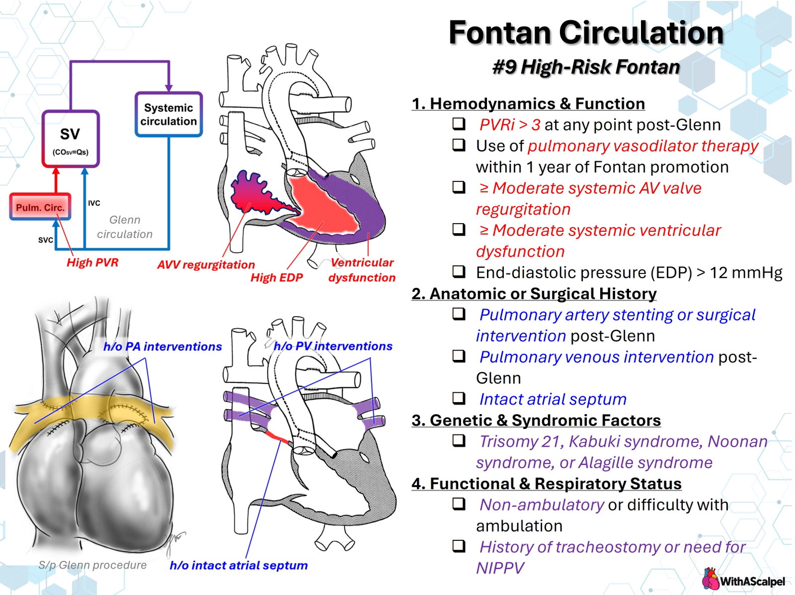
The accompanying illustration organizes these features into four domains: (1) hemodynamics and ventricular function, (2) anatomic or surgical history, (3) genetic and syndromic factors, and (4) functional and respiratory status. The principal objective of risk assessment is to distinguish correctable lesions from intrinsic physiology that may remain poorly tolerated after completion.
2. Hemodynamic Basis of Fontan Risk
2.1. Pulmonary vascular resistance
In a Glenn circulation, superior vena caval blood flows passively into the pulmonary arteries; after Fontan completion, inferior vena caval flow is added to this pathway. Total pulmonary blood flow becomes more dependent on low resistance across the entire pulmonary circuit. An indexed PVR (PVRi) above 3 Wood units·m² after Glenn palliation is a concerning feature in the supplied risk framework, but it is not a universally accepted absolute exclusion threshold.
PVRi is calculated from the transpulmonary pressure gradient divided by indexed pulmonary blood flow. Interpretation requires accurate estimates of effective pulmonary flow and pulmonary venous or atrial pressure. Aortopulmonary collaterals, antegrade pulmonary flow, uneven branch perfusion, and anesthetic conditions can substantially alter the calculation. A 2026 retrospective study of 81 patients proceeding to Fontan completion found that PVR estimated by combined cardiac magnetic resonance (CMR) flow and catheter-derived pressure measurements was associated with adverse events, whereas catheterization-only PVR was not discriminative [2]. This supports physiologically integrated measurement rather than reliance on an isolated threshold.
A historical elevation in PVRi after the Glenn operation matters, but current physiology and the response to treatment matter more than an isolated past value. Persistent elevation despite correction of obstruction and optimization of pulmonary vascular conditions raises concern for inadequate Fontan flow. Glenn-stage pulmonary arterial pressure and transpulmonary gradient have also been associated with subsequent outcomes, particularly in subsets with a dominant systemic right ventricle [3].
2.2. Ventricular end-diastolic pressure and systolic function
The systemic ventricle supplies the kinetic energy ultimately sustaining Fontan flow. Elevated ventricular end-diastolic pressure (EDP) raises pulmonary venous and left atrial pressure, diminishing the gradient available to drive transpulmonary flow. The illustration identifies EDP above 12 mmHg as a high-risk marker. This is a useful screening trigger, not a universally established operative cutoff; pressure must be interpreted alongside loading conditions, waveform quality, ventricular morphology, and atrioventricular (AV) valve function.
Importantly, a ventricle may have apparently preserved ejection fraction while exhibiting impaired compliance. Diastolic dysfunction can therefore compromise Fontan performance even without overt systolic dysfunction. Moderate or greater systemic ventricular systolic dysfunction further reduces cardiac reserve, particularly when postoperative preload falls. Contemporary analysis of Fontan failure identifies systolic dysfunction, elevated resistance, restrictive ventricular physiology, and obstructive lesions as distinct mechanisms of failure [4].
2.3. Systemic AV valve regurgitation
Moderate or severe systemic AV valve regurgitation creates an additional volume load and raises atrial pressure, often worsening pulmonary venous congestion and ventricular dilation. Regurgitation and ventricular dysfunction may amplify each other. A recent combined CMR–catheterization cohort associated moderate-to-severe AV valve regurgitation with adverse Fontan outcomes [2].
Assessment must distinguish valve morphology from functional regurgitation due to ventricular geometry or annular dilation. Repairability, durability of coaptation, and the possibility of postoperative stenosis all matter. The presence of significant regurgitation warrants consideration of intervention, but simultaneous valve repair at Fontan completion is not automatically preferable to earlier repair or further optimization.
2.4. Pulmonary vasodilator use
Pulmonary vasodilator therapy within the year preceding Fontan completion is included as a risk indicator in the illustration. It may reflect concern about elevated PVR, limited pulmonary arterial growth, or suboptimal Glenn hemodynamics. However, treatment exposure is a surrogate for underlying disease severity—not proof that medication itself increases risk. The drug, indication, response, and contemporaneous catheterization data should be reviewed before classifying the patient.
3. Anatomic and Surgical-History Risk Factors
3.1. Pulmonary artery interventions after Glenn
Previous pulmonary artery stenting, patch arterioplasty, or surgical reconstruction signals possible branch pulmonary artery narrowing, hypoplasia, asymmetric perfusion, or an unfavorable cavopulmonary connection. Even relatively localized stenosis may be important when pulmonary flow is passive. Imaging should assess the central confluence, Glenn anastomosis, branch origins, distal vascular beds, and prospective Fontan anastomotic geometry.
A history of intervention is not equivalent to residual disease. A widely patent, well-developed pulmonary arterial tree after successful reconstruction may carry substantially different physiological implications from recurrent obstruction. Risk depends on residual resistance and flow distribution, not merely on the number of procedures. Studies evaluating outcomes after stage-two palliation underscore the importance of assessing the entire interstage course rather than relying only on the most recent pre-Fontan measurement [5].
3.2. Pulmonary venous obstruction and prior intervention
Prior pulmonary vein intervention is particularly concerning because venous obstruction produces elevated downstream pulmonary pressures and can cause secondary pulmonary vascular remodeling. Stenosis may be recurrent, diffuse, or confined to individual veins, making mean atrial pressure an incomplete reflection of regional hemodynamics.
Pre-Fontan imaging should establish whether each pulmonary vein is patent, whether stenosis is progressive, and whether intervened segments have durable caliber. Recurrent multivessel disease may represent a much less modifiable risk than isolated branch pulmonary artery stenosis.
3.3. Intact or highly restrictive atrial septum
The slide includes a history of intact atrial septum. This should be understood primarily as a marker of the neonatal disease course, particularly in hypoplastic left heart syndrome, when restricted pulmonary venous egress may produce severe pulmonary venous hypertension and secondary pulmonary vascular or lymphatic injury.
A retrospective cohort of 191 Fontan patients reported early failure in eight (4%); neonatal balloon atrial septostomy was the only variable statistically associated with that outcome, although the small number of events limited interpretation [6]. This finding supports attention to the history of pulmonary venous decompression. It does not mean an atrial septum should remain intact or restrictive in a patient proceeding through single-ventricle palliation; adequate pulmonary venous egress is essential.
4. Genetic, Syndromic, and Extracardiac Factors
The illustration identifies trisomy 21, Kabuki syndrome, Noonan syndrome, and Alagille syndrome. Their relevance is not uniform and should not be interpreted as equivalent or absolute contraindications.
Trisomy 21 may coexist with pulmonary vascular disease, upper-airway obstruction, sleep-disordered breathing, and recurrent respiratory illness. A multicenter administrative-database study found increased mortality, prolonged ventilation, and resource utilization among patients with trisomy 21 undergoing cavopulmonary connections; residual confounding and selection effects remain important [7].
Noonan syndrome and other RASopathies can involve lymphatic dysplasia and myocardial disease. Alagille syndrome may involve peripheral pulmonary arterial abnormalities and clinically significant hepatobiliary disease. Kabuki syndrome can be accompanied by complex cardiac lesions, airway problems, immune dysfunction, and developmental impairment. For these less common syndromes, Fontan-specific outcome evidence is substantially more limited than for trisomy 21; decisions should be based on the patient's actual phenotype rather than a diagnostic label.
Hepatic disease, nutritional status, immune function, developmental needs, and lymphatic abnormalities should be considered alongside cardiac physiology. In particular, syndromic hepatic or lymphatic disease may complicate recognition and management of subsequent Fontan-associated organ dysfunction.
5. Functional and Respiratory Risk
Nonambulatory status or difficulty ambulating may indicate neuromuscular disease, developmental impairment, deconditioning, or substantial cardiac limitation. These etiologies have different prognostic implications. Activity level should therefore be evaluated in relation to baseline neurological and musculoskeletal function, rather than treated as a stand-alone hemodynamic measure.
A history of tracheostomy or a requirement for noninvasive positive-pressure ventilation (NIPPV) highlights potentially important airway and respiratory disease. Pulmonary blood flow in the Fontan circuit is sensitive to changes in intrathoracic pressure, lung volume, hypoxia, hypercapnia, and pulmonary vasoconstriction. Excessive mean airway pressure can impair systemic venous return and passive pulmonary flow. Conversely, effective airway support may reduce obstruction, hypoventilation, and secondary elevation of PVR. Thus, the need for NIPPV is a risk marker requiring individualized physiological assessment, not a reason to withdraw beneficial respiratory support.
Evaluation should identify fixed airway obstruction, sleep-disordered breathing, restrictive pulmonary mechanics, pulmonary parenchymal disease, aspiration, and the likelihood of prolonged postoperative ventilation.
6. Integrated Pre-Fontan Assessment
High-risk evaluation is most useful when it translates each abnormal finding into an actionable mechanism.
6.1. Echocardiography and cross-sectional imaging
Echocardiography should characterize systemic ventricular morphology and function, AV valve regurgitation, atrial communication, outflow tract obstruction, and accessible cavopulmonary pathways. CMR or CT can define branch pulmonary arteries, pulmonary veins, collateral burden, systemic venous anatomy, and spatial relationships important for Fontan construction. Combined CMR–catheterization has also been described as a practical method of integrating anatomic and hemodynamic evaluation in selected pre-Fontan patients [8].
6.2. Catheterization
When indicated, catheterization should provide mean pulmonary arterial and atrial pressures, ventricular EDP, cardiac output or flow estimates, transpulmonary gradient, and resistance. Measurements should be interpreted with oxygenation, sedation or ventilation, residual antegrade pulmonary blood flow, collateral circulation, and regional vascular anatomy in mind. If a measured elevation in PVR or filling pressure would change the decision to proceed, its reliability and reversibility deserve particular scrutiny.
6.3. Lymphatic assessment
High systemic venous pressure increases lymph formation and impedes central lymphatic drainage. Thoracic lymphatic abnormalities may therefore identify vulnerability not explained by conventional catheterization alone. In a retrospective MRI cohort, high-grade lymphatic abnormalities after Glenn were associated with adverse Fontan outcomes [9]. Another cohort of 238 Fontan completions found early complications in 58 patients (24%) under a broad composite definition; high-grade preoperative lymphatic imaging abnormalities were associated with approximately sixfold higher odds of complications [10]. Lymphatic imaging can strengthen assessment in selected patients, although universal screening and preventive interventions are not yet supported by definitive comparative evidence.
7. Operative Planning and Risk Modification
The first question is whether identified abnormalities can be corrected before completion. Potential interventions include relief of branch pulmonary artery stenosis, assessment or treatment of pulmonary venous obstruction, management of important AV valve regurgitation, reduction of collateral-related volume loading, and optimization of pulmonary vascular or respiratory conditions. Their sequence and expected benefit must be individualized.
At Fontan completion, the surgeon should establish a low-resistance connection with appropriate conduit orientation, no kinking, minimal distortion of the pulmonary arterial confluence, and unobstructed systemic and pulmonary venous pathways. Post-bypass assessment should evaluate Fontan pressure, atrial pressure, transpulmonary gradient, ventricular filling and output, AV valve competence, pulmonary artery and venous patency, and signs of maldistributed flow.
Fenestration provides a controlled right-to-left decompression pathway that may improve ventricular preload and reduce systemic venous pressure at the cost of systemic desaturation and potential paradoxical embolic risk. Evidence comparing routine and selective fenestration is heterogeneous; it should not be regarded as a complete solution for severe pulmonary vascular disease, important pulmonary venous obstruction, or markedly elevated ventricular EDP [11].
For particularly vulnerable patients, a planned postoperative strategy should anticipate low cardiac output, elevated venous pressure, pleural or chylous effusions, respiratory interactions, and the need for early catheter or surgical reassessment. Persistent hemodynamic compromise should prompt urgent evaluation for a correctable residual lesion rather than attribution to “Fontan physiology” alone.
8. Outcomes and Interpretation of Evidence
High-risk status indicates a spectrum, not an inevitable outcome. In a contemporary 398-patient extracardiac Fontan series, early mortality was 1.5%, and cumulative Fontan failure occurred in 12.5% over a median 10.3-year follow-up. Right ventricular dominance, aortic atresia, and elevated pulmonary artery pressure were associated with failure [12]. These outcomes describe a selected operative cohort and cannot be directly applied to patients who never reached Fontan completion.
In another retrospective cohort of 634 patients, later Fontan failure had multiple clinical manifestations and was associated with right ventricular dominance and higher pre-Fontan pulmonary artery pressure [13]. Definitions of “failure” differ markedly among studies, including early death or takedown, mechanical circulatory support, persistent effusions, protein-losing enteropathy, plastic bronchitis, and late transplantation. This heterogeneity prevents assigning one validated probability to every item in the illustration.
Selection bias is especially important: patients with the most unfavorable physiology may undergo additional interventions, have Fontan completion deferred, remain with Glenn circulation, or proceed to a different treatment pathway. Observational associations should therefore inform a multidisciplinary discussion rather than be converted into fixed prohibitions.
9. Key Clinical Principles
- Risk is multidimensional. PVR, ventricular EDP, AV valve competence, pulmonary arterial and venous anatomy, respiratory physiology, and extracardiac disease interact.
- Thresholds are screening markers. PVRi above 3 Wood units·m² and EDP above 12 mmHg warrant scrutiny but are not universally validated absolute contraindications.
- History and current physiology must be separated. Prior stenting, vasodilator use, or atrial septal intervention should trigger investigation of the underlying mechanism and its present severity.
- Pulmonary venous hypertension is particularly harmful. Venous obstruction, AV valve regurgitation, and ventricular stiffness all reduce the effective transpulmonary driving gradient.
- Syndromic risk is phenotype-dependent. Respiratory, lymphatic, hepatic, and developmental findings require individual assessment.
- Correction precedes completion when feasible. Fontan construction should follow deliberate evaluation of reversible lesions, with planned postoperative surveillance and early escalation if residual obstruction or low output is suspected.
References
- Rychik J, Atz AM, Celermajer DS, et al. Evaluation and Management of the Child and Adult With Fontan Circulation: A Scientific Statement From the American Heart Association. Circulation. 2019. DOI: 10.1161/CIR.0000000000000696. PMID: 31256636.
- Hsieh N, Doctor PN, Akam-Venkata J, et al. Elevated combined pulmonary vascular resistance obtained by cardiovascular magnetic resonance and cardiac catheterisation on pre-Fontan assessment is associated with adverse postoperative Fontan outcomes. Cardiol Young. 2026;36:62–67. DOI: 10.1017/S1047951125110664. PMID: 41555595.
- Vitanova K, Shiraishi S, Mayr B, et al. Impact of Characteristics at Stage-2-Palliation on Outcome Following Fontan Completion. Pediatr Cardiol. 2019;40:1476–1487. DOI: 10.1007/s00246-019-02172-6. PMID: 31342112.
- Van Puyvelde J, Rega F, Budts W, et al. Defining the causes for Fontan circulatory failure in total cavopulmonary connection patients. Interdiscip Cardiovasc Thorac Surg. 2024;39:ivae188. DOI: 10.1093/icvts/ivae188. PMID: 39565923.
- Ono M, Burri M, Mayr B, et al. Risk Factors for Failed Fontan Procedure After Stage 2 Palliation. Ann Thorac Surg. 2021;112:610–618. DOI: 10.1016/j.athoracsur.2020.06.030. PMID: 32828751.
- Rochelson E, Richmond ME, LaPar DJ, Torres A, Anderson BR. Identification of Risk Factors for Early Fontan Failure. Semin Thorac Cardiovasc Surg. 2020;32:522–528. DOI: 10.1053/j.semtcvs.2020.02.018. PMID: 32087242.
- Allen P, Anderson BR, Bacha E, LaPar DJ. Trisomy 21 Patients Undergoing Cavopulmonary Connections Need Improved Preoperative and Postoperative Care. Ann Thorac Surg. 2021;112:2012–2019. DOI: 10.1016/j.athoracsur.2020.10.014. PMID: 33161018.
- Eilers LF, Britt JJ, Weigand J, et al. Pre-Fontan Assessment Utilizing Combined Cardiac Catheterization and Cardiac MRI: Comparison to the Pre-Fontan Catheterization. Pediatr Cardiol. 2024;45:1448–1454. DOI: 10.1007/s00246-023-03178-x. PMID: 37210685.
- Biko DM, Dewitt AG, Pinto E, et al. MRI Evaluation of Lymphatic Abnormalities in the Neck and Thorax after Fontan Surgery: Relationship with Outcome. Radiology. 2019;291:774–780. DOI: 10.1148/radiol.2019180877. PMID: 30938628.
- Ghosh RM, Griffis HM, Glatz AC, et al. Prevalence and Cause of Early Fontan Complications: Does the Lymphatic Circulation Play a Role? J Am Heart Assoc. 2020;9:e015318. DOI: 10.1161/JAHA.119.015318. PMID: 32223393.
- Toncu A, Rădulescu CR, Dorobanţu D, Stoica S. Does routine fenestration improve early and late postoperative outcomes in patients undergoing Fontan palliation? Interact Cardiovasc Thorac Surg. 2020;30:773–779. DOI: 10.1093/icvts/ivaa002. PMID: 32243525.
- Ishigami S, King G, Buratto E, et al. Outcomes of extracardiac Fontan operation: A single institution experience with 398 patients. J Thorac Cardiovasc Surg. 2025;169:400–410.e7. DOI: 10.1016/j.jtcvs.2024.05.030. PMID: 38897544.
- Gaebert P, Schaeffer T, Palm J, et al. Incidence, pathophysiology, and treatment of failing Fontan after the total cavopulmonary connection. Cardiol Young. 2024;34:2406–2413. DOI: 10.1017/S1047951124025782. PMID: 39358848.