1. Concept and Clinical Significance
The Fontan circulation directs systemic venous blood to the pulmonary arteries without a subpulmonary ventricle. Pulmonary blood flow therefore depends on elevated systemic venous pressure and a relatively low-resistance pulmonary vascular bed. This arrangement sustains oxygenation but exposes the systemic organs and lymphatic system to chronic venous congestion. Lymphatic dysfunction is consequently an important mechanism of Fontan morbidity, not simply a secondary consequence of overt ventricular failure. Its principal manifestations include protein-losing enteropathy (PLE), plastic bronchitis, chylothorax, ascites, and peripheral edema [1].
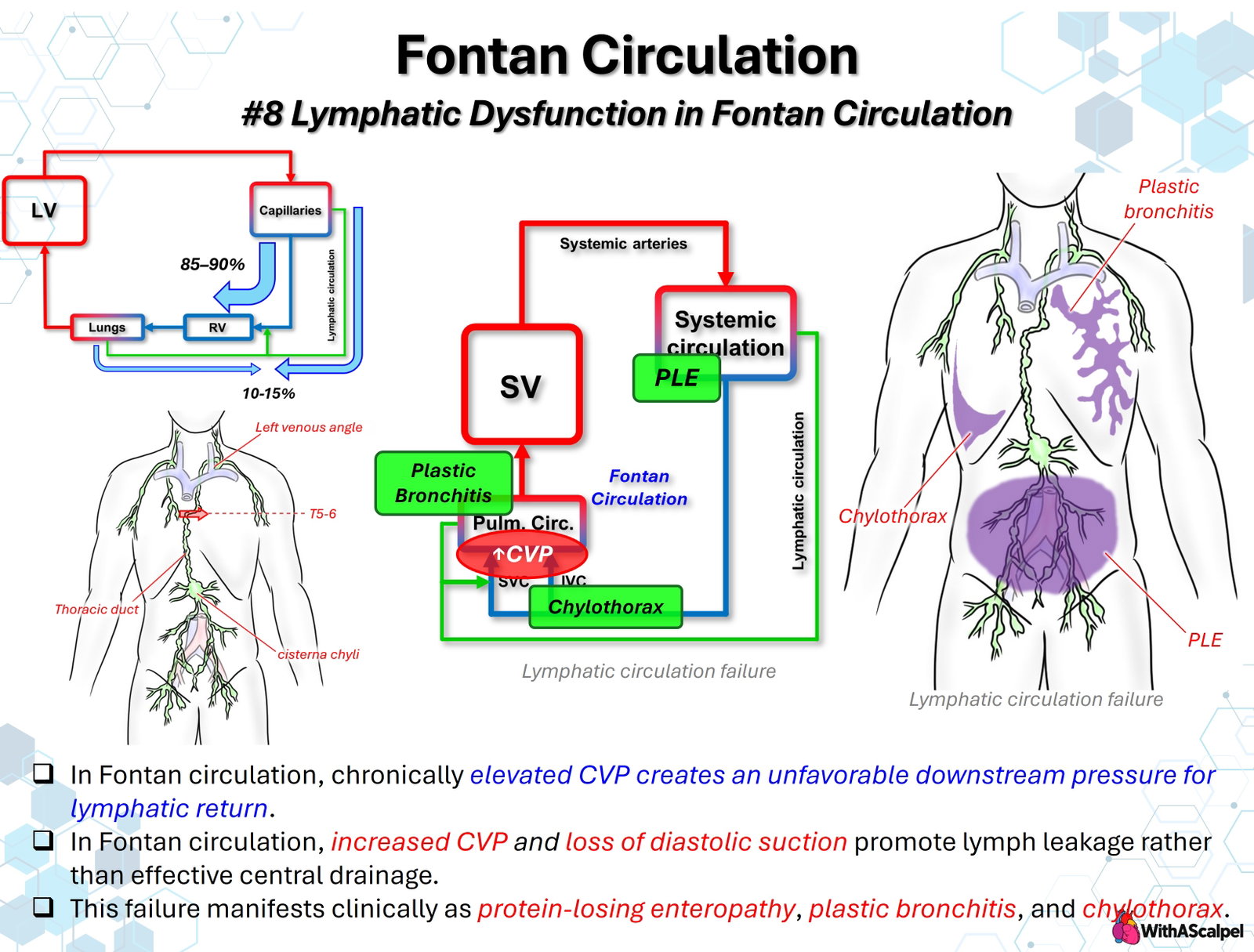
The accompanying illustration emphasizes a fundamental imbalance: increased lymph formation occurs simultaneously with impaired drainage into the systemic veins. When lymphatic transport capacity is exceeded, lymph can escape into the intestinal lumen, airways, or pleural space. These manifestations share an adverse hemodynamic substrate but arise from different anatomical lymphatic pathways; they require distinct diagnostic and therapeutic strategies.
2. Normal Lymphatic Anatomy and Physiology
2.1. Formation and central return of lymph
Fluid and plasma proteins continuously leave the microcirculation and enter interstitial tissues. Initial lymphatic capillaries collect this fluid, and larger collecting vessels transport it through lymph nodes toward the central venous circulation. The thoracic duct typically begins near the cisterna chyli in the upper abdomen, ascends through the posterior mediastinum, and terminates near the left internal jugular–subclavian venous junction. Its tributaries drain most of the lower body, abdominal viscera, and left upper thorax. The right lymphatic duct, when present as a distinct vessel, drains portions of the right upper body. Anatomical variation is common.
The liver and gastrointestinal tract are particularly important lymph-producing organs. Hepatic sinusoidal filtration generates protein-rich lymph, while intestinal lacteals carry absorbed long-chain dietary fats as chyle. The thoracic duct normally returns these fluids to the bloodstream against a small pressure difference at its venous outlet.
The slide's conventional depiction of 85–90% direct capillary reabsorption and 10–15% lymphatic return should not be interpreted as a fixed physiological partition. Under the revised Starling principle, sustained venous-end capillary reabsorption is generally limited, and lymphatic drainage is the principal pathway for returning filtered interstitial fluid and proteins to the circulation. This distinction strengthens, rather than weakens, the importance of lymphatic function.
2.2. Forces supporting lymphatic flow
Collecting lymphatics contain valves and smooth muscle that generate intrinsic phasic contractions. Respiratory pressure variations, skeletal muscle activity, and tissue movement provide additional propulsion. Forward flow ultimately depends on the balance between pressure generated within lymphatic vessels and the pressure at their venous outlet.
The low venous pressure of a normal biventricular circulation facilitates central lymph drainage. Classical hemodynamic observations demonstrated that systemic venous hypertension can simultaneously increase lymph production and obstruct its return into the veins [2]. More recent physiological studies in Fontan patients have documented altered lymphatic morphology and reduced pumping capacity [3].
3. Mechanisms of Lymphatic Failure in Fontan Circulation
3.1. Elevated downstream pressure
The thoracic duct ordinarily empties into a venous territory exposed to systemic venous pressure. After Glenn or Fontan palliation, this outlet is exposed to higher pressures than in normal circulation. The resulting reduction in the pressure gradient across the lymphovenous junction makes central lymphatic drainage less efficient.
In a retrospective study of 31 Fontan patients with lymphatic failure, Savla and colleagues found that higher central venous pressure was associated with thoracic duct dilatation. Larger ducts demonstrated a smaller pressure rise during temporary occlusion, suggesting impaired contractile reserve [4]. These physiological observations support the concept of lymphatic pump dysfunction, although they do not establish an individual pressure threshold at which failure becomes inevitable.
3.2. Increased lymph production and impaired transport
Venous hypertension elevates hydrostatic pressure in hepatic and splanchnic vascular beds, increasing capillary filtration and lymph production. Hepatic congestion may be especially important because the liver produces large quantities of protein-rich lymph. Meanwhile, elevated thoracic duct outlet pressure restricts effective return. The Fontan lymphatic system therefore faces both increased inflow and increased outflow resistance.
Reduced ventricular filling and cardiac output can worsen the problem indirectly by promoting neurohormonal activation and fluid retention. Pulmonary vascular obstruction, elevated pulmonary vascular resistance, atrioventricular valve regurgitation, ventricular diastolic dysfunction, or obstruction within the Fontan connection may further raise systemic venous pressure.
The phrase “loss of diastolic suction” in the illustration is best understood as the absence of a normal low-pressure, subpulmonary cardiac receiving compartment—not as complete loss of lymphatic propulsion. Intrinsic lymphatic contractions and respiration-dependent pressure changes persist after Fontan completion. Positive-pressure ventilation, particularly excessive intrathoracic pressure, may adversely affect both cavopulmonary flow and lymphatic return.
3.3. Abnormal lymphatic pathways and patient susceptibility
Not every patient with elevated Fontan pressure develops clinically significant lymphatic disease. Congenital lymphatic architecture, remodeling, collateral formation, impaired lymphatic contractility, and inflammatory or infectious triggers may help explain variable susceptibility.
The distribution of abnormal lymphatic channels is clinically decisive. Retrograde flow into peribronchial networks can produce plastic bronchitis; hepatoduodenal and intestinal lymphatic connections may cause PLE; and pleural lymphatic leakage can produce chylothorax. Severe thoracic lymphatic abnormalities identified before Fontan completion also carry prognostic information. In a retrospective MRI cohort of 83 patients, Biko and colleagues reported Fontan-completion failure in 54% of patients with type 4 lymphatic abnormalities versus 2% with type 1 or 2 abnormalities [5].
In a separate single-center cohort of 238 Fontan completions, 58 patients (24%) met a broad definition of early complications. Among those with available preoperative lymphatic MRI, high-grade abnormalities were associated with approximately sixfold greater odds of early complications [6]. These findings support lymphatic imaging for selected risk assessment but do not prove that preventive lymphatic intervention improves survival.
4. Major Clinical Manifestations
4.1. Protein-losing enteropathy
PLE is abnormal loss of protein-rich lymph into the gastrointestinal lumen. Abnormal hepatic and intestinal lymphatic channels can communicate with the duodenal wall, allowing lymph to enter the gut rather than return to the central venous circulation. Direct lymphangiographic evidence of hepatoduodenal leakage has been demonstrated in patients with congenital heart disease and elevated venous pressure [7].
Clinical findings include hypoalbuminemia, edema, ascites, diarrhea, impaired growth, fatigue, and malnutrition. Loss of immunoglobulins and lymphocytes can cause immune dysfunction; depletion of anticoagulant proteins and altered hemostasis may coexist with the underlying Fontan-associated thrombotic risk. A normal or mildly elevated Fontan pressure at a single catheterization does not exclude a clinically important lymphatic mechanism.
Diagnosis combines serum albumin and total protein assessment with evidence of gastrointestinal protein loss, most commonly an elevated fecal alpha-1-antitrypsin concentration or clearance. Renal protein loss, reduced hepatic synthesis, inadequate nutrition, and other causes of hypoalbuminemia must be considered. Persistent or refractory PLE warrants evaluation of Fontan hemodynamics and lymphatic anatomy.
4.2. Plastic bronchitis
Plastic bronchitis is characterized by formation of branching, often tenacious casts within the tracheobronchial tree. Abnormal pulmonary lymphatic perfusion allows lymphatic fluid and inflammatory or proteinaceous material to enter airways, where casts form. The principal clinical consequences are cough, wheezing, hypoxemia, recurrent respiratory distress, and potentially life-threatening airway obstruction.
Bronchoscopy establishes the extent of cast obstruction and can be immediately therapeutic. In a retrospective series of 18 patients with congenital heart disease and plastic bronchitis, Dori and colleagues identified retrograde pulmonary lymphatic flow in 16. Among 17 patients undergoing lymphatic intervention, 15 experienced substantial symptomatic improvement at reported follow-up [8]. Although influential, this experience was nonrandomized and cannot establish equivalent efficacy across all lymphatic phenotypes.
4.3. Chylothorax and related effusions
Chylothorax represents accumulation of chyle within the pleural cavity. It may follow direct lymphatic injury during surgery or reflect lymphatic hypertension, abnormal pleural perfusion, or impaired thoracic duct outflow. In Fontan patients, these mechanisms may coexist. Consequently, a postoperative chylous effusion does not automatically indicate a discrete surgically repairable thoracic duct injury.
Persistent chylothorax causes intravascular volume depletion, protein and immunoglobulin loss, lymphopenia, nutritional deficiencies, and respiratory compromise. Pleural fluid triglycerides and demonstration of chylomicrons, when necessary, help establish the diagnosis; fluid appearance alone is unreliable, particularly when enteral fat intake is limited. Related lymphatic failure may manifest as chylous ascites, nonchylous persistent effusions, or systemic edema [9].
5. Diagnostic Evaluation and Lymphatic Imaging
Initial assessment should distinguish hemodynamic drivers from the anatomical site of lymphatic leakage. Echocardiography evaluates ventricular performance, atrioventricular valve regurgitation, and obvious pathway obstruction. Cardiac MRI or CT can delineate cavopulmonary geometry, pulmonary arteries, pulmonary veins, and collateral vessels. Catheterization permits direct assessment of Fontan, branch pulmonary arterial, and pulmonary venous atrial pressures, together with cardiac output, resistance, and potential obstructive gradients. Rhythm disturbances and thrombosis also require evaluation.
Heavily T2-weighted MR lymphangiography depicts fluid-filled lymphatic structures without direct contrast injection. Thoracic imaging may be classified from type 1, with minimal abnormalities, through type 4, with extensive mediastinal and pulmonary lymphatic abnormalities [5]. This technique maps lymphatic burden but does not directly establish the direction of flow.
Dynamic contrast-enhanced MR lymphangiography (DCMRL) provides complementary functional information. Intranodal contrast injection delineates central lymphatic transit, thoracic duct drainage, and abnormal pulmonary or pleural pathways. Intrahepatic DCMRL can reveal hepatic lymphatic connections to the duodenum, particularly in PLE. A small initial series demonstrated the technical feasibility and clinical value of this targeted hepatic approach [10]. Intramesenteric imaging may provide additional information when intestinal lymphatic abnormalities remain incompletely characterized.
Imaging access and protocol should therefore match the suspected phenotype. A patient with recurrent airway casts may require detailed assessment of pulmonary lymphatic perfusion, whereas refractory PLE raises concern for hepatoduodenal or periduodenal channels.
6. Management: Relieve Congestion and Target the Leak
6.1. Optimize Fontan physiology
Treatment begins by addressing potentially reversible causes of venous hypertension and reduced forward flow. Relevant interventions include relief of Fontan or branch pulmonary arterial obstruction, treatment of arrhythmias, management of atrioventricular valve regurgitation, optimization of ventricular filling, and selected pulmonary vasodilator therapy when physiology supports its use. Diuresis may reduce congestion but should be balanced against the Fontan circulation's dependence on sufficient preload.
Fenestration creation or enlargement can decompress the Fontan pathway in selected patients, at the cost of increased right-to-left shunting and lower arterial oxygen saturation. It is neither a definitive treatment for every lymphatic complication nor an adequate substitute for correcting an anatomical obstruction.
6.2. Stabilize the affected organ system
For PLE, high-protein nutrition, judicious use of medium-chain triglycerides, diuretics, albumin replacement when clinically indicated, and replacement of deficient micronutrients or immunoglobulins may be appropriate. Medical strategies such as budesonide, heparin, or pulmonary vasodilators have been used in selected cases, but responses are inconsistent and supporting evidence is limited.
For plastic bronchitis, airway stabilization and bronchoscopic cast extraction take priority during acute obstruction. Airway clearance, inhaled mucolytic or fibrinolytic therapies, and treatment of superimposed inflammation may serve as adjuncts rather than correction of the underlying lymphatic lesion.
For chylothorax, pleural drainage when necessary, dietary reduction of long-chain fats with medium-chain triglyceride supplementation, and careful replacement of fluid, electrolytes, and proteins are common initial measures. Temporary bowel rest, parenteral nutrition, or octreotide may be considered according to clinical severity and local protocols. Prolonged drainage should prompt reassessment rather than indefinite conservative treatment.
6.3. Image-guided selective lymphatic intervention
Targeted embolization aims to interrupt an abnormal lymphatic communication while preserving useful central lymphatic drainage. Selective embolization of hepatoduodenal or peritracheal channels has produced symptomatic and biochemical improvement in small Fontan series [11].
More recent evidence illustrates both progress and limitations. In a 2026 single-center retrospective cohort of 71 patients with congenital heart disease and medically refractory PLE, combined intrahepatic and periduodenal embolization was associated with an initial response in 87% compared with 41% after isolated intrahepatic embolization. Sustained remission for at least one year occurred in 40% versus 11%, respectively. However, recurrent disease and procedure-related adverse events were substantial; the comparison was nonrandomized and included a high-risk, heterogeneous population [12].
Embolization should not be equated with indiscriminate thoracic duct sacrifice. Occlusion of central outflow may aggravate upstream lymphatic hypertension or redirect leakage into another compartment.
6.4. Thoracic duct decompression and surgical options
For severe, diffuse, or multicompartment lymphatic failure, thoracic duct decompression seeks to establish drainage into a lower-pressure receiving chamber. Surgical innominate vein turn-down or transcatheter approaches can redirect the thoracic duct-containing venous territory toward the pulmonary venous atrium. These procedures may reduce lymphatic afterload but introduce anatomical complexity, potential venous or lymphatic obstruction, and systemic desaturation from right-to-left venous shunting.
In a 12-patient transcatheter series, lymphatic failure resolved in six and improved in two at short-term follow-up [13]. A small surgical experience also demonstrated technical feasibility of concomitant lymphatic decompression in selected patients with severe pre-Fontan lymphatic abnormalities [14]. Neither approach has sufficient comparative evidence to justify routine use. Persistent failure despite optimized hemodynamics and specialized intervention should trigger multidisciplinary assessment for Fontan revision, other advanced support strategies, or transplantation.
7. Long-Term Surveillance and Prognosis
Lymphatic complications may arise shortly after Fontan completion or many years later. Follow-up should assess growth, nutritional status, albumin, immune function, fluid retention, exercise tolerance, recurrent respiratory symptoms, and the durability of previous lymphatic interventions. Serial lymphatic imaging is most informative when it addresses a defined clinical question rather than functioning as an isolated substitute for hemodynamic evaluation.
Importantly, clinical improvement after embolization or decompression does not necessarily mean that the underlying Fontan circulation has normalized. Recurrence, redistribution of lymphatic leakage, progressive hepatic congestion, and deterioration of cardiac output remain possible. Evidence for long-term durability comes predominantly from specialized centers and observational cohorts.
Key Clinical Principles
- Dual hemodynamic burden: Fontan venous hypertension increases lymph production while impairing central lymphatic return.
- Anatomy determines phenotype: Hepatoduodenal leakage causes PLE, pulmonary lymphatic leakage produces plastic bronchitis, and pleural leakage causes chylothorax.
- CVP is not sufficient: A single venous pressure measurement cannot define lymphatic function or identify the site of leakage.
- Image the flow pathway: T2-weighted MRI maps lymphatic burden; DCMRL identifies abnormal flow and intervention targets.
- Correct reversible hemodynamic lesions: Effective lymphatic treatment begins with assessment of the entire Fontan circuit.
- Preserve necessary drainage: Selective embolization and thoracic duct decompression address different mechanisms and are not interchangeable.
- Recognize the evidence limits: Promising interventional outcomes largely derive from small or nonrandomized cohorts; sustained follow-up remains essential.
References
- Rychik J, Atz AM, Celermajer DS, et al. Evaluation and Management of the Child and Adult With Fontan Circulation: A Scientific Statement From the American Heart Association. Circulation. 2019;140:e234–e284. DOI: 10.1161/CIR.0000000000000696. PMID: 31256636.
- Witte MH, Dumont AE, Clauss RH, Rader B, Levine N. Lymph Circulation in Congestive Heart Failure: Effect of External Thoracic Duct Drainage. Circulation. 1969;39:723–733. DOI: 10.1161/01.CIR.39.6.723. PMID: 5785287.
- Mohanakumar S, Telinius N, Kelly B, et al. Morphology and Function of the Lymphatic Vasculature in Patients With a Fontan Circulation. Circ Cardiovasc Imaging. 2019;12:e008074. DOI: 10.1161/CIRCIMAGING.118.008074. PMID: 30943769.
- Savla JJ, Kelly B, Krogh E, et al. Occlusion Pressure of the Thoracic Duct in Fontan Patients With Lymphatic Failure: Does Dilatation Challenge Contractility? World J Pediatr Congenit Heart Surg. 2022;13:737–744. DOI: 10.1177/21501351221119394. PMID: 36300263.
- Biko DM, DeWitt AG, Pinto EM, et al. MRI Evaluation of Lymphatic Abnormalities in the Neck and Thorax after Fontan Surgery: Relationship with Outcome. Radiology. 2019;291:774–780. DOI: 10.1148/radiol.2019180877. PMID: 30938628.
- Ghosh RM, Griffis HM, Glatz AC, et al. Prevalence and Cause of Early Fontan Complications: Does the Lymphatic Circulation Play a Role? J Am Heart Assoc. 2020;9:e015318. DOI: 10.1161/JAHA.119.015318. PMID: 32223393.
- Itkin M, Piccoli DA, Nadolski G, et al. Protein-Losing Enteropathy in Patients With Congenital Heart Disease. J Am Coll Cardiol. 2017;69:2929–2937. DOI: 10.1016/j.jacc.2017.04.023. PMID: 28619193.
- Dori Y, Keller MS, Rome JJ, et al. Percutaneous Lymphatic Embolization of Abnormal Pulmonary Lymphatic Flow as Treatment of Plastic Bronchitis in Patients With Congenital Heart Disease. Circulation. 2016;133:1160–1170. DOI: 10.1161/CIRCULATIONAHA.115.019710. PMID: 26864093.
- Tomasulo CE, Chen JM, Smith CL, Maeda K, Rome JJ, Dori Y. Lymphatic Disorders and Management in Patients With Congenital Heart Disease. Ann Thorac Surg. 2022;113:1101–1111. DOI: 10.1016/j.athoracsur.2020.10.058. PMID: 33373590.
- Biko DM, Smith CL, Otero HJ, et al. Intrahepatic dynamic contrast MR lymphangiography: initial experience with a new technique for the assessment of liver lymphatics. Eur Radiol. 2019;29:5190–5196. DOI: 10.1007/s00330-019-06112-z. PMID: 30887210.
- Maleux G, Storme E, Cools B, et al. Percutaneous embolization of lymphatic fistulae as treatment for protein-losing enteropathy and plastic bronchitis in patients with failing Fontan circulation. Catheter Cardiovasc Interv. 2019;94:996–1002. DOI: 10.1002/ccd.28501. PMID: 31596530.
- Smith CL, Gartenberg AJ, Ford B, et al. Outcomes After Lymphatic Embolization for Protein-Losing Enteropathy in Congenital Heart Disease. J Am Coll Cardiol. 2026;88:1473–1494. DOI: 10.1016/j.jacc.2026.07.036. PMID: 42644784.
- Smith CL, Dori Y, O'Byrne ML, Glatz AC, Gillespie MJ, Rome JJ. Transcatheter Thoracic Duct Decompression for Multicompartment Lymphatic Failure After Fontan Palliation. Circ Cardiovasc Interv. 2022;15:e011733. DOI: 10.1161/CIRCINTERVENTIONS.121.011733. PMID: 35708032.
- Kreutzer C, Klinger DA, Chiostri B, et al. Lymphatic Decompression Concomitant With Fontan/Kreutzer Procedure: Early Experience. World J Pediatr Congenit Heart Surg. 2020;11:284–292. DOI: 10.1177/2150135120905656. PMID: 32294012.