1. Concept and Clinical Significance
Pulmonary arteriovenous malformations (PAVMs) are abnormal pulmonary vascular pathways that allow blood to pass from the pulmonary arterial to the pulmonary venous circulation without normal alveolar–capillary gas exchange. This produces an intrapulmonary right-to-left physiological shunt, with desaturated blood returning to the pulmonary venous atrium and subsequently entering the systemic circulation. PAVMs range from discrete, angiographically visible fistulas to diffuse microscopic vascular abnormalities. The diffuse form is particularly important after cavopulmonary connection in patients with functionally univentricular hearts. [1]
The defining clinical manifestation is progressive systemic arterial desaturation despite anatomically patent cavopulmonary connections and adequate gross pulmonary blood flow. PAVMs may arise during the bidirectional Glenn stage, become prominent after a Kawashima procedure, or persist or develop following Fontan completion when hepatic venous blood is unevenly delivered to the lungs.
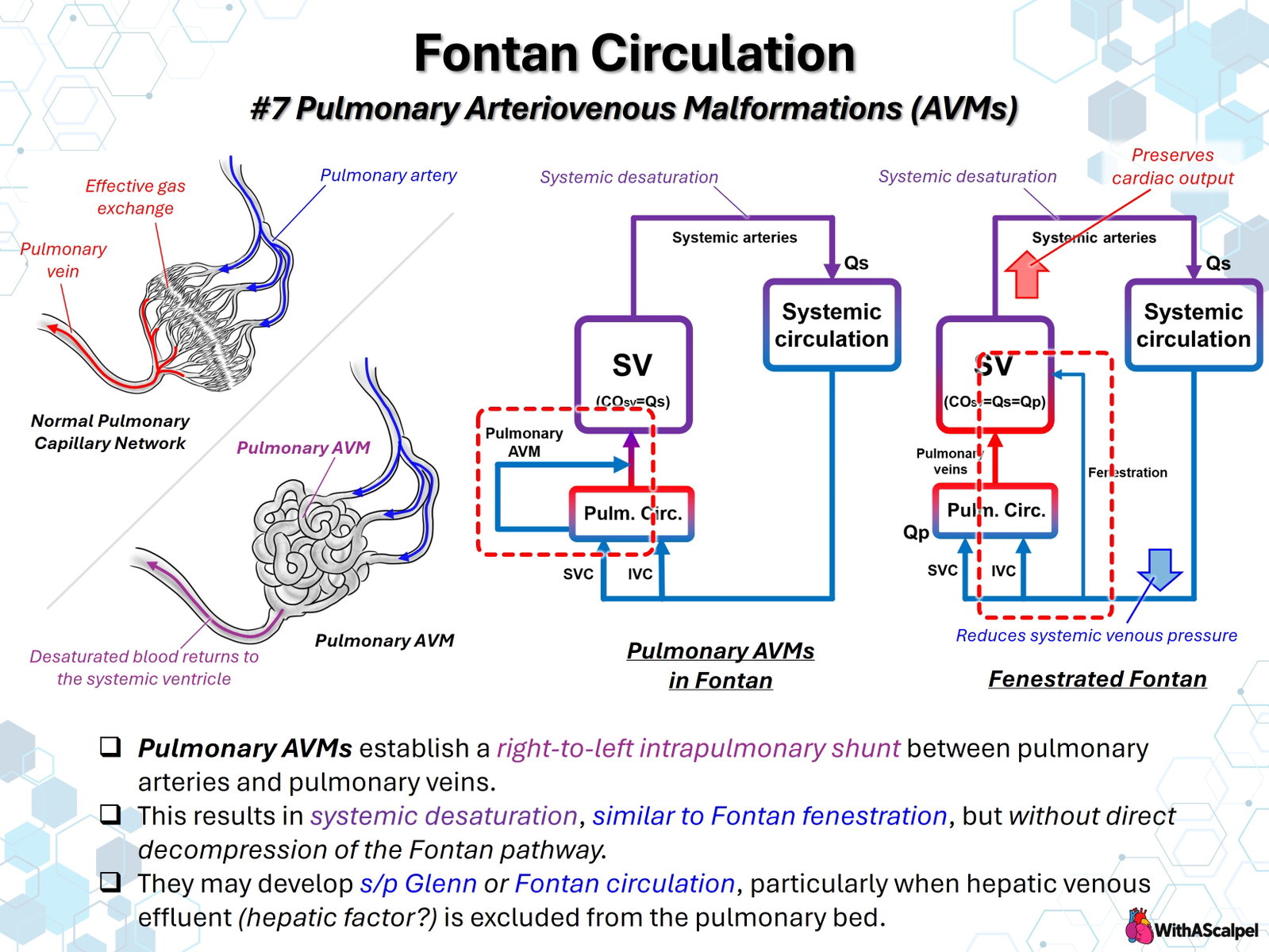
The central distinction in the accompanying illustration is that PAVMs reduce effective pulmonary oxygenation but do not directly decompress the Fontan pathway. In contrast, a Fontan fenestration creates an intentional communication from the high-pressure systemic venous pathway to the pulmonary venous atrium.
2. Pulmonary Microvascular Anatomy and Shunt Physiology
2.1. Normal versus abnormal pulmonary pathways
In normal lungs, pulmonary arterial blood enters progressively smaller arteries, arterioles, and the alveolar capillary network. This microvascular arrangement provides a large surface area and sufficient transit time for oxygen uptake and carbon dioxide elimination. Oxygenated blood then enters the pulmonary veins.
A PAVM provides a lower-resistance route that bypasses part or all of the effective alveolar capillary network. In diffuse disease, numerous abnormal microvascular channels and dilated vessels may be involved rather than a single large artery-to-vein connection. Blood passing through these pathways reaches the pulmonary veins with an oxygen content closer to mixed systemic venous blood.
The resulting desaturation depends on the fraction of pulmonary blood flow bypassing functional gas exchange, systemic venous oxygen content, hemoglobin concentration, and the oxygenation of blood traversing normally ventilated lung regions. Supplemental oxygen may increase saturation in blood passing through functional alveoli, but it cannot fully correct a substantial anatomical shunt.
2.2. Why PAVMs differ from Fontan fenestration
For an idealized nonfenestrated Fontan circulation, without significant collateral or residual shunt flow, total pulmonary arterial flow and effective systemic blood flow are approximately equal:
With PAVMs, blood still enters the pulmonary arterial circulation and subsequently returns through the pulmonary veins. Therefore, the presence of PAVMs alone does not require to become less than . Instead, the fraction of participating in effective gas exchange decreases. Here, refers to total pulmonary arterial flow, not just oxygenating microvascular flow.
With a Fontan fenestration, a separate portion of systemic venous return bypasses the pulmonary arteries and enters the pulmonary venous atrium:
A fenestration may lower systemic venous pressure and maintain ventricular preload and systemic cardiac output when pulmonary vascular resistance or atrial pressure is unfavorable. These benefits come at the expense of lower systemic arterial saturation. PAVMs can produce a similar saturation deficit without the same pressure-relief mechanism, because the shunt occurs downstream of the Fontan pathway within the pulmonary vascular bed. [2]
A low arterial saturation therefore cannot distinguish fenestration flow from PAVMs, venovenous collaterals, pulmonary parenchymal disease, or other residual right-to-left pathways. Their hemodynamic consequences and treatments are different.
3. Why PAVMs Develop After Cavopulmonary Connection
3.1. Hepatic venous effluent and the hepatic-factor hypothesis
The most persuasive clinical association is exclusion of hepatic venous effluent from the affected lung. In a landmark retrospective study of patients with congenital heart disease, Srivastava and colleagues identified 10 patients with PAVMs and reported a strong association between cavopulmonary connections that excluded normal hepatic venous blood and subsequent vascular malformations. Among a selected population with interrupted inferior vena cava and azygos continuation, PAVMs occurred in 6 of 28 patients with cavopulmonary connection compared with 1 of 56 without it. [3]
This observation gave rise to the hepatic-factor hypothesis: one or more circulating substances delivered through normal hepatic venous blood may suppress abnormal pulmonary vascular remodeling or maintain microvascular integrity. The responsible factor has not been conclusively identified. Experimental and histological investigations have implicated altered angiogenic signaling; for example, pulmonary tissue from children after cavopulmonary anastomosis demonstrated increased expression of vascular endothelial growth factor and its receptor in one small comparative study. These findings support biological plausibility but do not establish a single causal molecular pathway. [4]
Other proposed contributors include reduced pulmonary arterial pulsatility, chronic hypoxemia, abnormal shear stress, and regional differences in pulmonary perfusion. Their relative roles remain uncertain. Hepatic-flow exclusion is the strongest clinical association, but it should not be interpreted as proof that one identifiable circulating molecule completely explains PAVM formation.
3.2. Reversal after hepatic venous inclusion
Clinical regression after restoration of hepatic venous blood provides important evidence that at least some PAVMs are reversible. Shah and colleagues described three patients whose PAVMs resolved after hepatic veins were incorporated into the pulmonary circulation, with improvement occurring over months rather than immediately. [5]
The practical implication is that adequate hepatic venous delivery to both lungs—not merely a technically patent Fontan connection—is a relevant physiological objective. Structural regression and improved arterial saturation may lag behind successful reconstruction.
4. Patients and Anatomical Configurations at Risk
4.1. Glenn and Kawashima circulations
After a standard bidirectional Glenn procedure, superior caval blood enters the pulmonary arteries, while hepatic venous blood usually continues through the inferior caval–atrial pathway and is excluded from direct pulmonary arterial perfusion. PAVMs may consequently develop during prolonged Glenn physiology, especially when Fontan completion is delayed.
In interrupted inferior vena cava with azygos or hemiazygos continuation, a Kawashima connection directs most systemic venous return to the pulmonary arteries, while the hepatic veins may continue to drain separately into the atrium. This configuration is particularly susceptible to PAVMs because pulmonary arterial inflow can remain largely devoid of hepatic effluent. In 16 patients undergoing hepatic-vein incorporation for substantial PAVMs after Kawashima palliation, McElhinney and colleagues evaluated the clinical response to restoration of hepatic blood flow. [6]
In a later retrospective cohort of 22 patients after Kawashima operation, 15 (68%) developed PAVMs. Following hepatic venous incorporation in 18 patients, mean systemic saturation increased from 77% before incorporation to 95% at follow-up, although improvement was not generally immediate and some patients had persistent cyanosis. These results reflect a selected single-center cohort rather than the prevalence of PAVMs across all Glenn or Fontan patients. [7]
A 2025 retrospective series of 21 Kawashima patients documented PAVMs both before and after hepatic venous incorporation. All four patients with newly recognized PAVMs after incorporation had bilateral superior venae cavae and inadequate hepatic venous delivery to one lung. This reinforces the importance of bilateral flow distribution and continued surveillance. [8]
4.2. Persistent or unilateral PAVMs after Fontan completion
Fontan completion usually restores hepatic venous effluent to the pulmonary arterial circulation. Nevertheless, complex anatomy may allow hepatic flow to stream preferentially toward one lung while the other receives predominantly superior caval blood. Important configurations include heterotaxy, interrupted inferior vena cava, bilateral superior venae cavae, asymmetrical branch pulmonary arteries, and substantial spatial separation between hepatic conduit and caval inflows.
Conduit geometry matters. Patient-specific computational studies have shown that conduit offset, branch pulmonary artery flow split, and extracardiac versus intra-atrial configuration influence the distribution of inferior caval or hepatic blood. There is no reliably optimal conduit design for every anatomy. [9]
A 2023 MRI-based computational study of 18 Fontan patients found associations between modeled hepatic flow distribution and pulmonary venous flow, simulated pulmonary vascular resistance, and resting arterial saturation. These correlations support individualized assessment, although model-derived variables should not be interpreted as direct proof of causation or as universally validated intervention thresholds. [10]
5. Clinical Presentation and Diagnostic Evaluation
Progressive cyanosis is the principal clue. Exercise desaturation, reduced functional capacity, clubbing, or secondary erythrocytosis may occur in advanced cases. Right-to-left intrapulmonary passage also reduces normal pulmonary capillary filtration of embolic material; therefore, paradoxical embolic complications are biologically plausible, although their absolute incidence in Fontan-associated diffuse PAVMs is not well established. [1]
Evaluation should first exclude more common or directly treatable causes of desaturation: a patent fenestration, systemic venous-to-pulmonary venous collaterals, residual atrial communications, pulmonary artery or Fontan pathway obstruction, pulmonary venous abnormalities, and parenchymal lung disease.
Contrast echocardiography is a sensitive method for identifying intrapulmonary right-to-left passage. Appearance of injected microbubbles in the pulmonary venous atrium or systemic ventricle after pulmonary vascular transit supports intrapulmonary shunting. Injection site and timing require careful interpretation in complex Fontan anatomy, because fenestrations, venovenous collaterals, and preferential flow paths can confound the findings.
In a 14-patient study after cavopulmonary anastomosis, Chang and colleagues compared pulmonary angiography with branch-pulmonary-artery bubble contrast echocardiography. [11] In a separate study of 20 patients after modified Fontan operations, contrast echocardiography detected intrapulmonary shunting in 9 patients compared with 2 detected by angiography, demonstrating that angiographically occult diffuse disease may be clinically relevant. [12]
Cardiac catheterization assesses Fontan pressure, atrial and ventricular filling pressures, branch pulmonary artery anatomy, and alternative pathways for desaturation. Pulmonary angiography may demonstrate diffuse, finely branching vascularity with early pulmonary venous opacification or discrete fistulas. Selective injections can help localize unilateral disease.
CT angiography provides detailed visualization of macroscopic vascular connections but may fail to reveal microscopic PAVMs. Cardiovascular MRI, including phase-contrast flow imaging and selected four-dimensional flow or computational modeling, can assess pulmonary flow distribution, conduit geometry, and preferential hepatic streaming. Diagnostic tests are complementary rather than interchangeable.
6. Management and Reconstructive Strategy
6.1. Restore appropriate hepatic blood distribution
For Glenn- or Kawashima-associated diffuse PAVMs, the central treatment is to incorporate hepatic venous return into pulmonary arterial flow when anatomically and physiologically feasible. Conventional Fontan completion may be appropriate, but the strategy must consider ventricular function, pulmonary vascular resistance, atrioventricular valve competence, and overall Fontan suitability. Hepatic-flow incorporation is not a justification for proceeding with an otherwise unsuitable Fontan circulation.
When PAVMs persist after Fontan completion, reconstructive planning should address preferential streaming. Potential strategies include repositioning the hepatic conduit, revising a superior cavopulmonary anastomosis, addressing branch pulmonary artery obstruction, creating a bifurcated hepatic conduit, or connecting the hepatic venous pathway to the azygos system to improve mixing.
In a retrospective study of eight patients with heterotaxy, unilateral hepatic streaming, and clinically significant PAVMs after Fontan completion, McElhinney and colleagues reported improvement in four of five patients who underwent hepatic-to-azygos connection; outcomes after other revisions were less consistent. This is valuable but small, nonrandomized surgical experience. [13]
Patient-specific virtual surgical planning has been applied to five patients with interrupted inferior vena cava and severe PAVMs, demonstrating how alternative configurations may produce markedly different hepatic flow distributions. Modeling helps compare geometry before intervention but does not replace clinical judgment or postoperative physiological validation. [14]
A published transcatheter case demonstrated creation of a hepatic conduit–azygos connection with improved oxygenation, supporting a catheter-based option in highly selected anatomy. Such complex interventions remain specialized, case-dependent approaches rather than standard first-line therapy. [15]
6.2. Why routine PAVM embolization is usually inappropriate
Embolization is an established treatment for selected discrete, accessible PAVMs, especially those unrelated to cavopulmonary physiology. However, Glenn- or Fontan-associated PAVMs are often numerous and diffuse. Occluding individual microscopic pathways does not correct the underlying absence or maldistribution of hepatic venous effluent and may be impractical or ineffective. The dominant therapeutic target is therefore the cavopulmonary circulation and its hepatic flow distribution, not routine coil occlusion of every visible abnormal vessel. [1]
6.3. Assessment after intervention
Following reconstruction, the essential assessments include unobstructed hepatic and superior caval pathways, balanced branch pulmonary artery perfusion, appropriate pulmonary and atrial pressures, preserved systemic output, and the absence of unintended residual shunts. A technically successful reconstruction may not immediately normalize saturation because regression of established PAVMs can require months.
7. Long-Term Surveillance
Serial arterial oxygen saturation should be assessed at rest and, when clinically informative, during exercise. Echocardiography evaluates ventricular function, atrioventricular valve regurgitation, pathway patency, and other causes of cyanosis. Persistent or worsening desaturation should prompt a targeted evaluation for PAVMs and competing shunt pathways rather than an assumption that the malformations are irreversible.
In complex Fontan anatomy, surveillance must consider whether hepatic venous flow actually reaches both lungs. Recurrent unilateral disease may indicate changing pulmonary flow splits, branch pulmonary artery narrowing, conduit geometry, or competitive superior caval streaming. Decisions about reintervention should incorporate anatomical findings, oxygen delivery, functional status, Fontan pressure, and anticipated procedural risk.
Key Clinical and Surgical Principles
- PAVMs cause intrapulmonary right-to-left shunting by bypassing effective alveolar–capillary oxygen exchange.
- PAVMs and Fontan fenestration are not hemodynamically equivalent: both can cause cyanosis, but only the fenestration directly bypasses and decompresses the Fontan venous pathway.
- Exclusion or maldistribution of hepatic venous blood is the dominant clinically supported association; the precise hepatic factor remains unidentified.
- Fontan completion is not sufficient by itself if hepatic blood preferentially streams to only one lung.
- Contrast echocardiography can reveal diffuse disease that conventional angiography does not clearly demonstrate.
- Reconstruct the source of abnormal streaming rather than routinely embolizing diffuse postoperative PAVMs.
- Allow time for regression and reassess systemic oxygenation and bilateral pulmonary flow distribution longitudinally.
References
[1] Cartin-Ceba R, Swanson KL, Krowka MJ. Pulmonary arteriovenous malformations. Chest. 2013;144:1033–1044. DOI: 10.1378/chest.12-0924. PMID: 24008954.
[2] Bridges ND, Lock JE, Castañeda AR. Baffle fenestration with subsequent transcatheter closure. Modification of the Fontan operation for patients at increased risk. Circulation. 1990;82:1681–1689. DOI: 10.1161/01.CIR.82.5.1681. PMID: 2225370.
[3] Srivastava D, Preminger T, Lock JE, et al. Hepatic venous blood and the development of pulmonary arteriovenous malformations in congenital heart disease. Circulation. 1995;92:1217–1222. DOI: 10.1161/01.CIR.92.5.1217. PMID: 7648668.
[4] Starnes SL, Duncan BW, Kneebone JM, et al. Angiogenic proteins in the lungs of children after cavopulmonary anastomosis. J Thorac Cardiovasc Surg. 2001;122:518–523. DOI: 10.1067/mtc.2001.115423. PMID: 11547304.
[5] Shah MJ, Rychik J, Fogel MA, Murphy JD, Jacobs ML. Pulmonary AV malformations after superior cavopulmonary connection: resolution after inclusion of hepatic veins in the pulmonary circulation. Ann Thorac Surg. 1997;63:960–963. DOI: 10.1016/S0003-4975(96)00961-7. PMID: 9124971.
[6] McElhinney DB, Kreutzer J, Lang P, Mayer JE Jr, del Nido PJ, Lock JE. Incorporation of the hepatic veins into the cavopulmonary circulation in patients with heterotaxy and pulmonary arteriovenous malformations after a Kawashima procedure. Ann Thorac Surg. 2005;80:1597–1603. DOI: 10.1016/j.athoracsur.2005.05.101. PMID: 16242423.
[7] Alsoufi B, Rosenblum J, Travers C, et al. Outcomes of single ventricle patients undergoing the Kawashima procedure: can we do better? World J Pediatr Congenit Heart Surg. 2019;10:20–27. DOI: 10.1177/2150135118809082. PMID: 30799723.
[8] Behrend L, Matsubara M, Osawa T, et al. Clinical outcomes of Kawashima procedure and subsequent hepatic vein incorporation. Eur J Cardiothorac Surg. 2025;67:ezaf058. DOI: 10.1093/ejcts/ezaf058. PMID: 39992217.
[9] Dasi LP, Whitehead K, Pekkan K, et al. Pulmonary hepatic flow distribution in total cavopulmonary connections: extracardiac versus intracardiac. J Thorac Cardiovasc Surg. 2011;141:207–214. DOI: 10.1016/j.jtcvs.2010.06.009. PMID: 20621314.
[10] Frieberg P, Sjöberg P, Hedström E, Carlsson M, Liuba P. In vivo hepatic flow distribution by computational fluid dynamics can predict pulmonary flow distribution in patients with Fontan circulation. Sci Rep. 2023;13:18206. DOI: 10.1038/s41598-023-45396-6. PMID: 37875552.
[11] Chang RK, Alejos JC, Atkinson D, et al. Bubble contrast echocardiography in detecting pulmonary arteriovenous shunting in children with univentricular heart after cavopulmonary anastomosis. J Am Coll Cardiol. 1999;33:2052–2058. DOI: 10.1016/S0735-1097(99)00096-0. PMID: 10362213.
[12] Larsson ES, Solymar L, Eriksson BO, de Wahl Granelli A, Mellander M. Bubble contrast echocardiography in detecting pulmonary arteriovenous malformations after modified Fontan operations. Cardiol Young. 2001. DOI: 10.1017/S1047951101000737. PMID: 11727905.
[13] McElhinney DB, Marx GR, Marshall AC, Mayer JE, del Nido PJ. Cavopulmonary pathway modification in patients with heterotaxy and newly diagnosed or persistent pulmonary arteriovenous malformations after a modified Fontan operation. J Thorac Cardiovasc Surg. 2011;141:1362–1370.e1. DOI: 10.1016/j.jtcvs.2010.08.088. PMID: 21146835.
[14] de Zélicourt DA, Haggerty CM, Sundareswaran KS, et al. Individualized computer-based surgical planning to address pulmonary arteriovenous malformations in patients with a single ventricle with an interrupted inferior vena cava and azygous continuation. J Thorac Cardiovasc Surg. 2011;141:1170–1177. DOI: 10.1016/j.jtcvs.2010.11.032. PMID: 21334010.
[15] Ratnayaka K, Wei ZA, Ryan JR, et al. Transcatheter hepatic conduit-azygous vein connection reduces pulmonary arteriovenous malformations in a cyanotic Fontan patient. JACC Case Rep. 2023;10:101760. DOI: 10.1016/j.jaccas.2023.101760. PMID: 36974056.