1. Concept and Clinical Significance
Systemic-to-pulmonary collateral arteries, commonly termed aortopulmonary collaterals (APCs), are acquired arterial connections that carry blood from the systemic arterial circulation into the pulmonary vascular bed. They are particularly relevant after bidirectional cavopulmonary connection and following Fontan completion, when pulmonary perfusion is intended to depend predominantly on passive systemic venous flow.
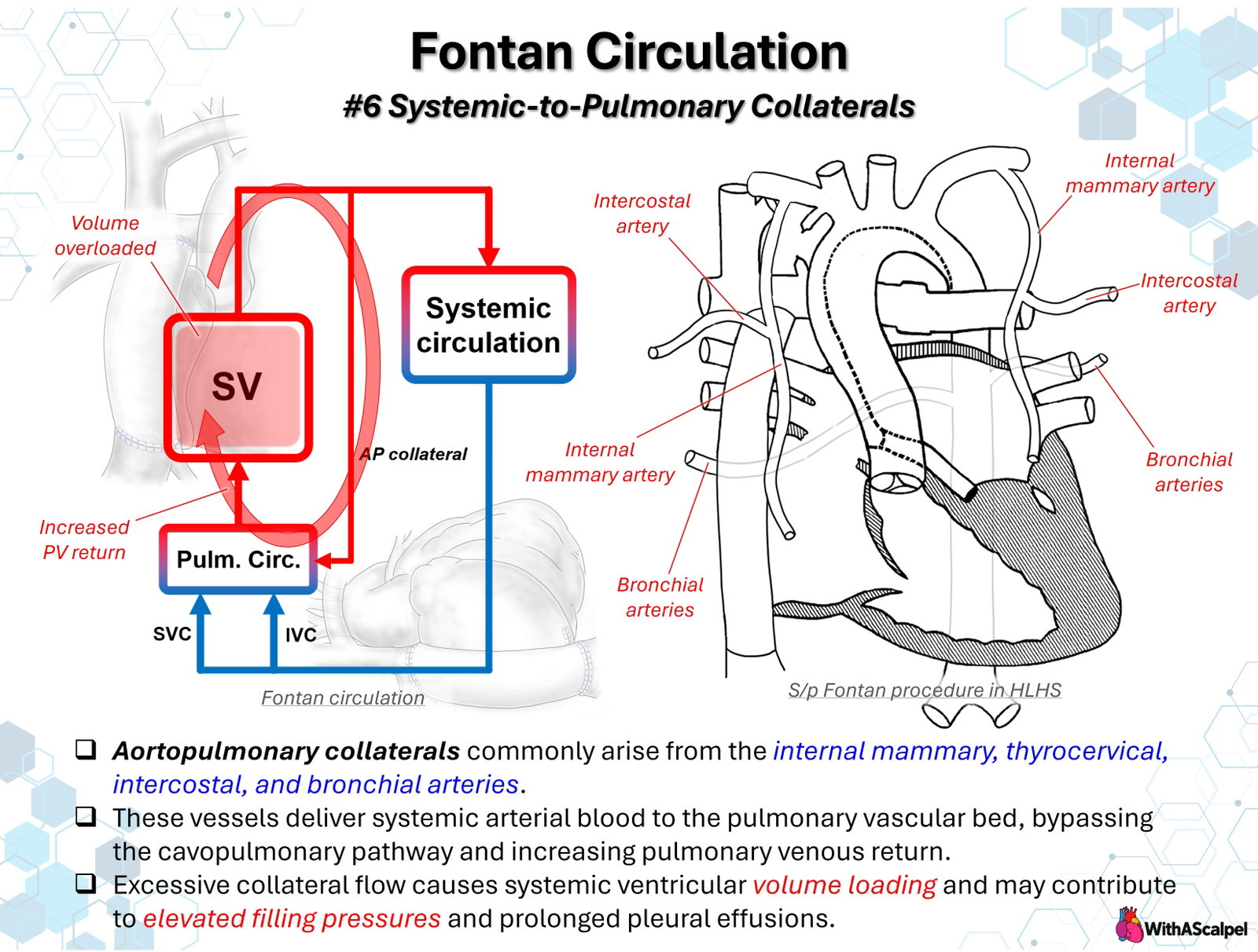
APCs create an additional systemic-to-pulmonary pathway that bypasses the cavopulmonary connection. Blood entering the lungs through these vessels returns through the pulmonary veins to the single ventricle and is ejected into the aorta, where a proportion may recirculate through the collateral network. Consequently, collateral flow can increase pulmonary venous return and systemic ventricular volume loading without necessarily increasing effective systemic tissue perfusion.
In a retrospective cohort of 430 patients undergoing staged single-ventricle palliation, angiographically identified APCs were present in 13% at the Glenn stage and 42% before Fontan completion. These figures reflect the specific cohort and angiographic detection method, rather than a universal prevalence. [1]
2. Vascular Anatomy and Collateral Formation
2.1. Sites of origin and pulmonary connections
APCs most often arise from branches of the subclavian arteries or descending thoracic aorta. Important sources include:
- Internal thoracic (internal mammary) arteries, coursing along the inner anterior chest wall and supplying pleural or mediastinal branches that may extend toward the lungs.
- Intercostal arteries, especially branches from the descending thoracic aorta that develop transpleural connections.
- Bronchial arteries, whose preexisting systemic bronchial circulation may enlarge and communicate with the pulmonary circulation.
- Thyrocervical and costocervical branches, together with other systemic arterial branches that can contribute to complex collateral networks.
The accompanying diagram illustrates several of these sources around the thoracic aorta, lungs, and reconstructed great vessels. Rather than consisting of a single discrete shunt, APCs may form tortuous, branching networks with multiple origins, anastomoses, and peripheral connections. Their distribution may be asymmetric and can be influenced by previous surgical shunts, thoracotomy, pleural adhesions, and regional pulmonary arterial supply.
An important anatomical distinction is that the usual APCs associated with staged Fontan palliation are not synonymous with major aortopulmonary collateral arteries (MAPCAs) in pulmonary atresia with ventricular septal defect. MAPCAs may provide essential segmental pulmonary blood supply and require a different anatomical assessment and reconstructive strategy. A vessel should never be considered expendable solely because it arises from the systemic arterial tree.
2.2. Mechanisms of development
The biological mechanisms are incompletely defined. Chronic cyanosis, altered pulmonary perfusion, regional hypoperfusion, previous systemic-to-pulmonary shunts, and angiogenic remodeling are plausible contributors. After a Glenn operation, reduced or heterogeneous antegrade pulmonary blood flow may favor enlargement of preexisting bronchial and pleural arterial channels.
Collateral flow varies substantially among patients and may persist after Fontan completion. Phase-contrast cardiovascular magnetic resonance (CMR) studies have demonstrated that these vessels can supply a substantial fraction of total pulmonary venous flow. [2] Smaller branch pulmonary arteries have also been associated with greater collateral flow and larger systemic ventricular volumes, although these associations do not establish that pulmonary artery hypoplasia directly causes collateral formation. [3]
3. Hemodynamics: A Recirculating Volume Load
3.1. Flow relationships
In an uncomplicated, nonfenestrated Fontan circulation without important collateral shunts, pulmonary arterial blood flow approximates systemic venous return, and pulmonary venous return approximates aortic output. APCs disrupt that correspondence.
In a simplified steady-state model:
Here, QAPC represents systemic arterial collateral flow, QPV total pulmonary venous return, and QAo aortic flow. These equations assume no other important shunts and adequate measurement of the relevant vessels. A Fontan fenestration, venovenous collaterals, residual surgical shunts, anomalous venous connections, or significant valvar regurgitation require a more complete flow accounting.
The key implication is that aortic cardiac output is not identical to effective systemic tissue flow when a substantial fraction is diverted through APCs. The ventricle may eject an apparently adequate volume while a meaningful proportion repeatedly circulates through the pulmonary bed and returns to the ventricle.
3.2. Effects on ventricular filling and pulmonary circulation
APCs increase pulmonary venous return and therefore ventricular preload. With a compliant ventricle and modest collateral burden, this may be tolerated. With progressive collateral flow, the resulting loading may enlarge the single ventricle, increase myocardial work, and aggravate atrioventricular valve regurgitation. When ventricular compliance is impaired, additional preload may raise atrial and ventricular filling pressures without a proportional benefit in effective forward systemic flow.
In the Fontan circulation, pulmonary blood flow depends on a limited pressure gradient between the systemic venous pathway and pulmonary venous atrium. An increase in pulmonary venous or atrial pressure can reduce this gradient and further compromise passive cavopulmonary flow. Collateral vessels may also increase regional pulmonary vascular flow and pressure. However, APCs do not invariably produce elevated Fontan pressure, and a high central venous pressure should not automatically be attributed to collateral flow.
3.3. Quantitative physiological evidence
In a 44-patient pre-Fontan CMR cohort, systemic-to-pulmonary collateral flow averaged 1.5 ± 0.9 L/min/m², equivalent to approximately 31% of aortic flow and 44% of pulmonary venous flow. Greater flow was associated with longer postoperative hospitalization and chest tube drainage. [4]
A separate prospective multimodality study similarly found that collateral flow could constitute a substantial component of pulmonary perfusion and was associated with longer postoperative pleural drainage and hospitalization. [5] The numerical values describe selected pre-Fontan cohorts, not an expected collateral burden in every established Fontan circulation.
4. Clinical Consequences
4.1. Ventricular volume overload
The defining ventricular consequence is recirculating volume loading. Serial echocardiography or CMR may show ventricular dilation, increasing atrioventricular valve regurgitation, or eventually elevated filling pressures. The functional impact depends on myocardial reserve, ventricular morphology, valvar competence, and the magnitude of collateral runoff. Ventricular dilation alone is not proof that APCs are the sole cause; valve regurgitation, residual shunts, and ventricular dysfunction must be assessed simultaneously. [3]
4.2. Pleural effusions and early Fontan recovery
Excess pulmonary blood flow and elevated pulmonary venous pressures can contribute to pulmonary and pleural fluid accumulation. Increased Fontan venous pressure may also impair lymphatic drainage. Thus, APCs are a plausible additional burden in patients with prolonged pleural effusions after Fontan completion, particularly when ventricular filling pressures or pulmonary vascular pressures are unfavorable.
An early retrospective series of 71 Fontan patients found that angiographically prominent APCs were associated with prolonged pleural drainage, and that collateral occlusion was associated with shorter drainage duration. [6] Subsequent CMR studies demonstrated similar associations between measured collateral flow and postoperative recovery. [4,5] These findings are predominantly observational and cannot establish that embolization will shorten effusions in every patient.
Persistent effusions require a broader evaluation for Fontan-pathway or branch pulmonary artery obstruction, elevated pulmonary vascular resistance, restrictive ventricular physiology, atrioventricular valve regurgitation, diaphragmatic dysfunction, and lymphatic abnormalities. APCs may be one contributor rather than the principal lesion.
4.3. Other considerations
Large collateral networks can create additional challenges during reoperation or transplantation because of enlarged systemic arterial branches and potential bleeding. In patients with low systemic oxygen saturation before Fontan completion, collateral perfusion may also support pulmonary oxygen uptake; abrupt occlusion may reduce saturation if native pulmonary arterial flow is inadequate. Therefore, the physiological significance of each vessel must be interpreted in the context of the stage of palliation and its pulmonary supply.
5. Diagnostic Evaluation and Flow Quantification
5.1. Echocardiography and CT angiography
Echocardiography evaluates the consequences of collateral loading—ventricular size and function, atrioventricular valve regurgitation, pulmonary venous pathway, and potential residual lesions—but generally cannot quantify the total collateral network reliably.
Contrast-enhanced CT angiography can delineate the origin, course, caliber, and pulmonary distribution of individual systemic arterial feeders. It is particularly useful for planning catheter access, identifying vessels near the mediastinum or chest wall, and assessing complex postoperative anatomy. CT provides vascular morphology rather than a direct measurement of aggregate collateral flow.
5.2. CMR: quantifying total collateral burden
Phase-contrast CMR or suitably acquired four-dimensional flow CMR can quantify the net systemic-to-pulmonary collateral contribution by comparing measured inflow and outflow:
Alternatively, in an appropriate anatomy:
These independent estimates serve as useful internal checks. CMR can report indexed collateral flow (L/min/m²), the proportion of aortic flow diverted through APCs, and the proportion of pulmonary venous return attributable to collateral flow. Contemporary Society for Cardiovascular Magnetic Resonance guidance explicitly includes collateral flow accounting in congenital single-ventricle examinations. [7]
Measurements require attention to plane positioning, arrhythmia, flow aliasing, small venous channels, fenestration location, and other shunts. A single angiographic estimate of collateral prominence is not interchangeable with quantified CMR flow.
5.3. Catheterization
Catheterization is especially valuable when intervention is being considered. Selective injection of internal thoracic, subclavian, bronchial, or intercostal branches identifies dominant feeders and their pulmonary connections. Simultaneously, invasive measurement of Fontan pressure, atrial or wedge pressure, transpulmonary gradient, and ventricular end-diastolic pressure helps determine whether collateral flow is an isolated problem or part of a more complex hemodynamic failure.
Angiography may underestimate diffuse microvascular collateral networks or fail to represent total flow because opacification depends on injection technique and vascular territory. A combined anatomical and physiological assessment is therefore preferable to treating every visible vessel.
6. Management and Embolization Strategy
6.1. When to consider treatment
Routine embolization of every APC is not supported by consistent clinical-outcome evidence. Selection should consider symptoms, objective collateral burden, systemic ventricular dilation or progressive atrioventricular valve regurgitation, elevated filling pressures, and otherwise unexplained or persistent pleural drainage. Significant vessels may also warrant attention in preparation for major cardiac reoperation or transplantation.
Before Fontan completion, decisions must account for the extent to which the lungs depend on collateral perfusion and whether substantial pulmonary artery obstruction is present. After Fontan completion, intervention is generally most compelling when a significant collateral burden plausibly contributes to ventricular loading or symptomatic circulatory inefficiency.
6.2. Transcatheter embolization
The usual intervention is selective transcatheter embolization with detachable coils, vascular plugs, or other embolic materials chosen according to vessel size and anatomy. Procedural planning should:
- Identify the major arterial feeders and demonstrate their pulmonary distribution.
- Evaluate whether the target vessel supplies essential lung tissue or gives rise to spinal, cerebral, coronary, or other critical branches.
- Select a stable embolization site while preserving normal systemic arterial branches whenever feasible.
- Confirm occlusion and reassess residual feeders, hemodynamics, and systemic oxygenation.
Intercostal and bronchial arterial procedures require particular caution because of potential spinal arterial connections. Non-target embolization, device migration, vascular injury, and contrast exposure are additional considerations. Diffuse networks may require staged, selective treatment rather than an attempt at complete anatomical eradication.
Surgical ligation may occasionally be feasible when large collateral vessels are encountered during operative exposure, but diffuse vessels cannot be reliably controlled through a single operative field. Surgical control should be individualized rather than treated as a substitute for careful preoperative vascular mapping.
7. Evidence, Controversies, and Contemporary Guidance
In the multicenter Pediatric Heart Network Fontan Cross-Sectional Study, 80 patients who had undergone pre-Fontan APC coiling were compared with 459 who had not. After adjustment, coiling was not associated with a shorter postoperative hospital stay or better late outcomes, while institutional coiling rates varied substantially. The study lacked standardized measurements of collateral burden and was subject to selection bias. [8]
A later single-center analysis of 227 Fontan patients examined pulmonary artery size, pre-Fontan collateral embolization timing, and postoperative pleural drainage, again emphasizing heterogeneity in the relationship between collateral intervention and effusions. [9] These investigations explain why a physiologically convincing rationale for occlusion has not translated into a universal mandate for prophylactic embolization.
The 2025 multisociety adult congenital heart disease guideline, published in 2026, states that closure can be useful in symptomatic adults with Fontan circulation or those with systemic ventricular volume overload (Class IIa, Level C–EO). [10] This is an adult-specific recommendation based largely on expert opinion, not proof that routine embolization benefits all pediatric pre-Fontan patients.
The most defensible approach is therefore individualized: measure or estimate collateral burden, establish its hemodynamic relevance, identify competing lesions, and intervene when the expected benefit exceeds the risks.
8. Key Clinical Principles
- APCs are a systemic-to-pulmonary recirculation pathway: they bypass the Fontan venous pathway and increase pulmonary venous return.
- Aortic output may overestimate effective systemic flow: some ventricular output is diverted back to the lungs through collateral vessels.
- Ventricular volume overload is central: excessive flow may increase ventricular dimensions, filling pressure, and atrioventricular valve regurgitation.
- Effusions are multifactorial: collateral burden is one possible contributor, not a sufficient explanation for persistent pleural drainage.
- CMR quantifies flow; CT and catheter angiography map anatomy: these modalities provide complementary information.
- Treat significant physiology, not every visible vessel: routine prophylactic embolization is not established by consistent outcome evidence.
- Protect critical arterial branches: selective embolization requires particular attention to spinal arterial connections.
References
- Schmiel M, Kido T, Georgiev S, et al. Aortopulmonary collaterals in single ventricle: incidence, associated factors and clinical significance. Interact Cardiovasc Thorac Surg. 2022;35(2):ivac190. DOI: 10.1093/icvts/ivac190. PMID: 35876534.
- Grosse-Wortmann L, Al-Otay A, Yoo SJ. Aortopulmonary collaterals after bidirectional cavopulmonary connection or Fontan completion: quantification with MRI. Circ Cardiovasc Imaging. 2009;2(3):219–225. DOI: 10.1161/CIRCIMAGING.108.834192. PMID: 19808596.
- Latus H, Gummel K, Diederichs T, et al. Aortopulmonary collateral flow is related to pulmonary artery size and affects ventricular dimensions in patients after the Fontan procedure. PLoS One. 2013;8(11):e81684. DOI: 10.1371/journal.pone.0081684. PMID: 24303064.
- Glatz AC, Rome JJ, Small AJ, et al. Systemic-to-pulmonary collateral flow, as measured by cardiac magnetic resonance imaging, is associated with acute post-Fontan clinical outcomes. Circ Cardiovasc Imaging. 2012;5(2):218–225. DOI: 10.1161/CIRCIMAGING.111.966986. PMID: 22228054.
- Grosse-Wortmann L, Drolet C, Dragulescu A, et al. Aortopulmonary collateral flow volume affects early postoperative outcome after Fontan completion: a multimodality study. J Thorac Cardiovasc Surg. 2012;144(6):1329–1336. DOI: 10.1016/j.jtcvs.2012.03.032. PMID: 22502974.
- Spicer RL, Uzark KC, Moore JW, Mainwaring RD, Lamberti JJ. Aortopulmonary collateral vessels and prolonged pleural effusions after modified Fontan procedures. Am Heart J. 1996;131(6):1164–1168. DOI: 10.1016/S0002-8703(96)90092-7. PMID: 8644596.
- Grosse-Wortmann L, Wald RM, Valverde I, et al. Society for Cardiovascular Magnetic Resonance guidelines for reporting cardiovascular magnetic resonance examinations in patients with congenital heart disease. J Cardiovasc Magn Reson. 2024;26(2):101062. DOI: 10.1016/j.jocmr.2024.101062. PMID: 39053855.
- Banka P, Sleeper LA, Atz AM, et al. Practice variability and outcomes of coil embolization of aortopulmonary collaterals before Fontan completion: a report from the Pediatric Heart Network Fontan Cross-Sectional Study. Am Heart J. 2011;162(1):125–130. DOI: 10.1016/j.ahj.2011.03.021. PMID: 21742098.
- Nabeshima T, Ishikawa Y, Sumitomo N, et al. The impact of the pulmonary artery index and aortopulmonary collateral artery coil embolization on intractable pleural effusions after a Fontan surgery. Int Heart J. 2021;62(3):559–565. DOI: 10.1536/ihj.20-498. PMID: 33994500.
- Gurvitz M, Krieger EV, Fuller S, et al. 2025 ACC/AHA/HRS/ISACHD/SCAI guideline for the management of adults with congenital heart disease: a report of the American College of Cardiology/American Heart Association Joint Committee on Clinical Practice Guidelines. J Am Coll Cardiol. 2026;87(7):822–976. DOI: 10.1016/j.jacc.2025.09.006. PMID: 41411480.