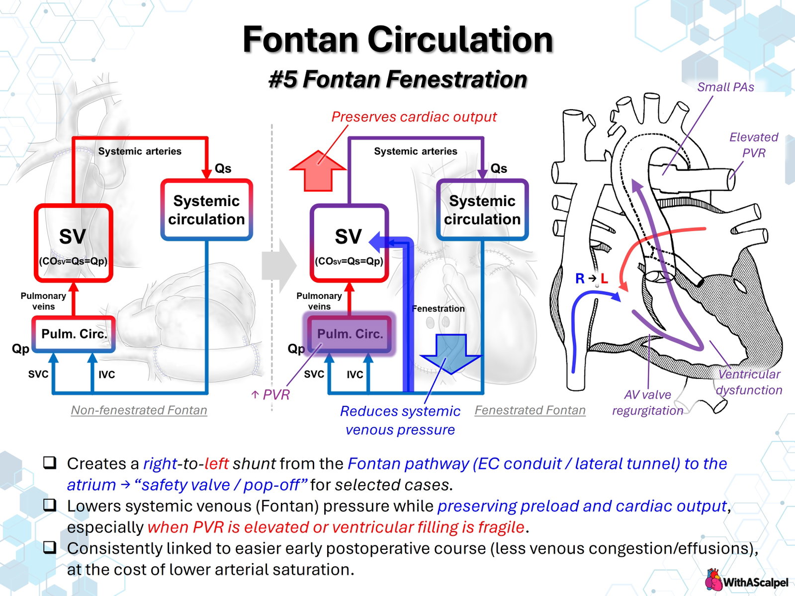
1. Concept and Physiological Rationale
Fontan fenestration is a deliberately created communication between the systemic venous Fontan pathway and the pulmonary venous atrium. It provides a controlled right-to-left shunt that bypasses the pulmonary vascular bed, allowing some systemic venous return to reach the single ventricle without passing through the pulmonary circulation. Its purpose is not to improve arterial oxygen saturation but to reduce the hemodynamic penalty of a circulation without a subpulmonary ventricle.
In a nonfenestrated Fontan circulation, both superior and inferior caval venous return must traverse the pulmonary arteries before reaching the systemic ventricle. Pulmonary blood flow depends on the pressure gradient between the Fontan pathway and pulmonary venous atrium, together with pulmonary vascular resistance (PVR) and other sources of pathway resistance. Even modest increases in resistance or ventricular filling pressure may produce systemic venous hypertension, reduced ventricular preload, and low cardiac output.
A fenestration acts as a pressure-relief pathway, traditionally called a “pop-off.” The concept was established in high-risk Fontan patients with ventricular dysfunction, elevated pulmonary artery pressure, atrioventricular (AV) valve regurgitation, or pulmonary artery distortion.[1] The benefit is conditional: a communication is useful when the reduction in venous congestion and preservation of forward systemic flow outweigh the accompanying arterial desaturation.
2. Hemodynamics: Why a Right-to-Left Shunt May Preserve Output
2.1. Flow distribution and pressure gradients
A patent fenestration creates two pathways from the systemic venous circuit: one through the pulmonary arteries and another directly into the pulmonary venous atrium. In a simplified circulation without significant collaterals, total systemic flow is the sum of pulmonary flow and fenestration flow:
Therefore, and are not necessarily equal in a fenestrated Fontan. This distinction matters when interpreting the diagram, systemic oxygen saturation, cardiac output, and catheterization measurements. In contrast, a nonfenestrated Fontan without collateral shunts has approximately equal systemic and pulmonary flow.
Fenestration flow depends primarily on the pressure difference between the Fontan pathway and atrium, the effective orifice area, and the dynamic resistance of the opening. When Fontan pressure rises relative to atrial pressure, right-to-left flow generally increases. This decompresses the upstream venous compartment and supplements ventricular filling through a pathway that avoids the pulmonary vascular resistance.
The resulting decrease in Fontan pressure may facilitate systemic venous and lymphatic drainage. At the same time, reduced dependence on transpulmonary flow can protect systemic output during periods of elevated PVR or impaired ventricular compliance. It does not correct pulmonary artery obstruction, elevated ventricular end-diastolic pressure, or AV valve regurgitation.
2.2. Oxygen saturation versus oxygen delivery
The principal physiological cost is admixture of systemic venous blood with pulmonary venous blood. Consequently, arterial oxygen saturation (SaO₂) falls as the right-to-left shunt increases. However, lower saturation does not automatically imply lower systemic oxygen delivery.
Systemic oxygen delivery is determined by:
where arterial oxygen content depends predominantly on hemoglobin concentration and saturation. If fenestration increases systemic flow sufficiently, oxygen delivery may be maintained or improved despite reduced SaO₂. Conversely, excessive shunting can lower arterial oxygen content without producing a clinically meaningful increase in flow. The relevant endpoint is perfusion and oxygen delivery, not saturation alone.
3. Anatomical Configuration and Surgical Creation
In a lateral tunnel Fontan, the communication is usually created through the intra-atrial baffle into the pulmonary venous atrium. Its position should allow a predictable atrial-level shunt without compromising caval flow or adjacent atrial structures.
In an extracardiac conduit Fontan, the conduit lies outside the atrium, so a communication must be constructed between the conduit and the pulmonary venous atrium. Techniques include a direct conduit-to-atrial opening or a short interposed tissue connection. The choice depends on conduit position, distance from the atrium, atrial wall mobility, and the ability to maintain a patent, unobstructed communication.[2]
Technical planning should address:
- Location: provide direct drainage to the pulmonary venous atrium while avoiding pulmonary venous inflow obstruction, the AV valve, and neighboring vascular structures.
- Orifice geometry: avoid an opening that is so small that it closes early or so large that excessive cyanosis results. The optimal diameter is patient- and strategy-dependent; no universal size is supported by comparative evidence.
- Conduit integrity: preserve smooth caval flow and avoid kinking, narrowing, or distortion of the extracardiac pathway.
- Hemostasis and patency: ensure secure tissue approximation and document the direction and magnitude of shunting on intraoperative echocardiography.
Surgical creation is generally planned during Fontan completion, but late surgical or transcatheter creation or enlargement may be considered as rescue therapy in selected patients with Fontan circulatory failure. Fenestration should not substitute for correction of a surgically or catheterically treatable obstruction.
4. Selecting Patients for Fenestration
There is no universally accepted mandate for routine fenestration. Practices range from routine creation to selective or infrequent use, reflecting differences in patient risk, operative technique, and institutional experience.
Fenestration is particularly reasonable to consider when the Fontan circulation is expected to have limited hemodynamic reserve:
- Elevated preoperative pulmonary artery pressure or PVR, including uncertain pulmonary vascular reactivity.
- Small or distorted branch pulmonary arteries, provided important obstructions are also addressed.
- Impaired systolic function or restrictive ventricular diastolic physiology, which limits pulmonary venous-to-ventricular filling.
- Moderate or greater systemic AV valve regurgitation or other factors increasing atrial and ventricular filling pressures.
- Borderline Fontan hemodynamics, substantial systemic-to-pulmonary collateral burden, or difficult early separation from cardiopulmonary bypass.
These are risk considerations rather than validated stand-alone indications. A retrospective study of 183 extracardiac Fontan patients found less chest drainage among fenestrated high-risk patients but not among lower-risk patients, illustrating possible effect modification by baseline physiology.[3] A patient with markedly unfavorable PVR, severe ventricular dysfunction, or uncorrected pathway obstruction may remain unsuitable for Fontan completion despite a planned fenestration.
5. Evidence for Early Postoperative Benefit
The strongest randomized evidence comes from a single-center trial of 49 standard-risk patients. Lemler and colleagues assigned 25 patients to fenestrated Fontan and 24 to nonfenestrated Fontan. Fenestration was associated with less chest tube drainage, a shorter hospital stay, and fewer additional postoperative procedures.[4] Although influential, this trial was small and reflects operative and perioperative practices of its era.
A 2020 meta-analysis pooled 19 studies involving 4,806 patients. Fenestration was associated with less need for prolonged pleural drainage and lower postoperative pulmonary artery pressure, as well as an expected decrease in oxygen saturation of approximately three percentage points. The pooled data did not demonstrate significant differences in early Fontan failure, death, or hospital length of stay.[5] Most included comparisons were observational, with heterogeneous risk profiles and technical approaches.
The educational statement that fenestration is “consistently linked to an easier postoperative course” therefore requires qualification. Reduction in venous congestion and pleural drainage is biologically plausible and supported by some clinical studies, but routine fenestration has not been proven to improve all major early outcomes. The decision should be individualized rather than inferred from saturation or Fontan pressure alone.
6. Early Postoperative and Intensive Care Implications
Immediately after Fontan completion, fenestration may become particularly valuable when PVR increases because of hypoxia, atelectasis, hypercarbia, acidosis, elevated intrathoracic pressure, or pulmonary vascular disease. It can maintain atrial filling and systemic output despite temporary resistance to forward pulmonary flow.
Assessment must integrate systemic venous pressure, atrial pressure when available, arterial blood pressure, lactate, urine output, near-infrared spectroscopy, mixed or systemic venous oxygen saturation, echocardiography, and the trend in chest drainage. The presence of a fenestration changes the interpretation of each variable.
Low saturation with preserved perfusion may reflect effective decompression rather than a primary pulmonary problem. Conversely, progressive desaturation with falling blood pressure, increasing lactate, or declining venous saturation suggests impaired oxygen delivery and requires urgent assessment. Excessive right-to-left flow can coexist with pulmonary artery stenosis, rising PVR, conduit obstruction, or ventricular dysfunction.
Management begins with identifying and correcting the cause of high Fontan resistance or elevated filling pressure. Optimize lung recruitment without unnecessarily high mean airway pressure, avoid hypoxia and acidosis, maintain appropriate intravascular volume, and address AV valve dysfunction, ventricular dysfunction, and anatomic obstruction. Extubation and spontaneous breathing may favor passive pulmonary blood flow in selected stable patients, but respiratory support should be guided by the overall cardiopulmonary condition.
Persistent pleural effusions or chylothorax warrant evaluation of Fontan pressures, conduit and branch pulmonary artery patency, pulmonary venous obstruction, AV valve function, and lymphatic abnormalities. A patent fenestration may reduce lymphatic afterload; it cannot independently resolve every cause of postoperative fluid accumulation.
7. Long-Term Consequences and Trade-offs
A patent fenestration can remain an important decompression route, particularly when Fontan physiology deteriorates. Nevertheless, chronic right-to-left shunting produces persistent cyanosis and may contribute to erythrocytosis, reduced exercise oxygen saturation, and potential paradoxical embolization. Long-term anticoagulation or antiplatelet decisions must consider overall Fontan-associated thrombotic risk, rhythm, prior thrombosis, ventricular function, and institutional protocols rather than fenestration status alone.
In the Pediatric Heart Network cross-sectional cohort of 536 Fontan patients, those with an open fenestration had lower resting saturation (median 89% versus 95%), without demonstrable differences in exercise performance, echocardiographic function, growth, or observed stroke/thrombosis prevalence.[6] A 2024 multicenter retrospective study of 407 patients found no statistically significant association between persistent fenestration and a composite of mortality, transplantation, atrial arrhythmia, or thromboembolism after adjustment; residual confounding and uncertainty regarding spontaneous closure remained important limitations.[7]
More recent data are not entirely uniform. In a 2026 four-center retrospective cohort of 105 previously fenestrated patients, a patent fenestration at one year was associated with a lower observed frequency of protein-losing enteropathy, but not a difference in Fontan-associated liver disease; patency groups differed in ventricular filling pressures.[8] These findings are hypothesis-generating, not proof that maintaining a fenestration prevents protein-losing enteropathy. Similarly, observations after closure in initially high-risk patients suggest that venovenous collateral formation may provide an alternative decompression route when Fontan physiology remains unfavorable.[9]
8. Evaluation and Closure of a Persistent Fenestration
A fenestration should not be closed solely to normalize pulse oximetry. The key question is whether the patient can tolerate redirecting the entire venous return through the pulmonary circulation without unacceptable increases in venous pressure or loss of systemic output.
Before closure, evaluate the Fontan pathway, branch pulmonary arteries, pulmonary veins, ventricular systolic and diastolic performance, AV valve regurgitation, aortopulmonary and venovenous collaterals, and any clinical evidence of Fontan-associated organ dysfunction. Catheterization is particularly useful when hemodynamics are uncertain or device closure is planned.
Temporary balloon test occlusion allows simultaneous assessment of arterial saturation, systemic venous pressure, atrial pressure, and cardiac index. In a classic 60-patient study, unfavorable responses were associated with greater venous hypertension and impaired systemic venous oxygenation, emphasizing the importance of identifying correctable lesions before closure.[10]
No threshold is universally validated. One published institutional protocol required an unobstructed pathway, baseline Fontan pressure at or below 15 mmHg, cardiac index at least 2 L/min/m², and no more than a 20% decrease in cardiac index during test occlusion.[11] These are institutional selection criteria, not a guideline or universal closure rule.
Device closure can durably improve SaO₂ in carefully selected patients. In a retrospective series of 51 closures, median saturation increased from 87% to 95%, while measured cardiac index decreased from 4.0 to 2.9 L/min/m² immediately after closure.[12] This illustrates why improved saturation and preserved output must be assessed together. Closure may be deferred in patients who depend on fenestration flow for hemodynamic stability.
9. Key Clinical Principles
- A Fontan fenestration is a controlled right-to-left bypass, intended to decompress systemic venous pressure and preserve systemic ventricular filling.
- Systemic flow can exceed pulmonary flow when fenestration is patent: .
- Lower SaO₂ is an expected trade-off, not necessarily evidence of lower systemic oxygen delivery.
- Benefit is physiology-dependent, particularly in patients with elevated PVR, limited ventricular compliance, AV valve regurgitation, or early postoperative instability.
- Evidence supports selected early benefits, especially reduced pleural drainage, but does not establish universal survival or hospital-stay benefit.
- A fenestration is not a substitute for correcting anatomic obstruction, ventricular dysfunction, or important AV valve regurgitation.
- Closure requires physiological testing, not saturation alone; test occlusion should demonstrate acceptable venous pressure and systemic output.
References
- Bridges ND, Lock JE, Castañeda AR. Baffle fenestration with subsequent transcatheter closure. Modification of the Fontan operation for patients at increased risk. Circulation. 1990;82:1681–1689. DOI: 10.1161/01.CIR.82.5.1681. PMID: 2225370.
- Do-Nguyen CC, Kilcoyne MF, Gray P, Jonas RA. The evolution of surgical technique of the fenestrated Fontan procedure. J Card Surg. 2020;35:1407–1409. DOI: 10.1111/jocs.14617. PMID: 32445189.
- Fan F, Liu Z, Li S, et al. Effect of fenestration on early postoperative outcome in extracardiac Fontan patients with different risk levels. Pediatr Cardiol. 2017;38:643–649. DOI: 10.1007/s00246-016-1561-x. PMID: 28116475.
- Lemler MS, Scott WA, Leonard SR, Stromberg D, Ramaciotti C. Fenestration improves clinical outcome of the Fontan procedure: a prospective, randomized study. Circulation. 2002;105:207–212. DOI: 10.1161/hc0202.102237. PMID: 11790702.
- Bouhout I, Ben-Ali W, Khalaf D, Raboisson MJ, Poirier N. Effect of fenestration on Fontan procedure outcomes: a meta-analysis and review. Ann Thorac Surg. 2020;109:1467–1474. DOI: 10.1016/j.athoracsur.2019.12.020. PMID: 31987825.
- Atz AM, Travison TG, McCrindle BW, et al. Late status of Fontan patients with persistent surgical fenestration. J Am Coll Cardiol. 2011;57:2437–2443. DOI: 10.1016/j.jacc.2011.01.031. PMID: 21658565.
- Dib N, Samuel M, Levesque S, et al. Impact of Fontan fenestration on adverse cardiovascular outcomes: a multicentre study. Can J Cardiol. 2024;40:1606–1615. DOI: 10.1016/j.cjca.2024.01.031. PMID: 38309467.
- Horie S, Shikata F, Oka N, et al. Impact of fenestration patency on long-term Fontan outcomes. Eur J Cardiothorac Surg. 2026;68:ezag047. DOI: 10.1093/ejcts/ezag047. PMID: 41557473.
- Ozawa H, Hoashi T, Ohuchi H, Kurosaki K, Ichikawa H. Long-term outcomes after fenestration closure in high-risk Fontan candidates. Pediatr Cardiol. 2021;42:1356–1364. DOI: 10.1007/s00246-021-02619-9. PMID: 33881601.
- Bridges ND, Lock JE, Mayer JE Jr, Burnett J, Castañeda AR. Cardiac catheterization and test occlusion of the interatrial communication after the fenestrated Fontan operation. J Am Coll Cardiol. 1995;25:1712–1717. DOI: 10.1016/0735-1097(95)00055-9. PMID: 7759728.
- Thatte N, Dimas V, Nugent A, et al. Use of institutional criteria for transcatheter device closure of Fontan fenestration—midterm outcomes. Ann Pediatr Cardiol. 2020;13:327–333. DOI: 10.4103/apc.APC_154_19. PMID: 33311921.
- Devanagondi R, Leonard GT. Transcatheter Fontan fenestration closure: sustained improvements in oxygen saturation with minimal morbidity and mortality. Pediatr Cardiol. 2023;44:922–926. DOI: 10.1007/s00246-022-03077-7. PMID: 36565311.