1. Overview
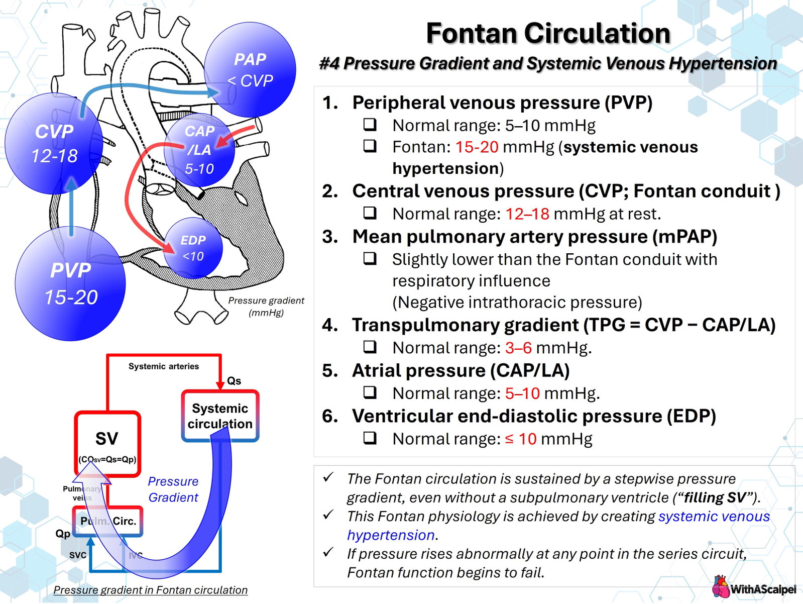
The Fontan circulation maintains pulmonary blood flow without a subpulmonary ventricle. Systemic venous return enters the pulmonary arteries through a total cavopulmonary connection (TCPC), traverses the pulmonary vascular bed, and reaches the pulmonary venous atrium before filling the systemic ventricle. Consequently, the circulation depends on a small pressure gradient sustained by elevated systemic venous pressure rather than the pulsatile pressure generated by a right ventricle. This arrangement produces the characteristic Fontan paradox: obligatory upstream venous congestion combined with restricted downstream ventricular preload.[1–3]
The clinical objective is not to maximize central venous pressure (CVP). It is to maintain sufficient pulmonary blood flow and systemic cardiac output at the lowest feasible Fontan pressure. Understanding the pressures at each point in the circuit is essential for interpreting catheterization data, identifying residual obstruction, and managing postoperative circulatory dysfunction.
2. Pressure Hierarchy Across the Fontan Circuit
In an unobstructed Fontan circulation, blood ordinarily moves down a series of mean pressure gradients:
Peripheral and central systemic veins → Fontan pathway → pulmonary arteries → pulmonary microcirculation → pulmonary venous atrium → systemic ventricle.
Unlike a biventricular circulation, there is no right ventricular systolic pressure rise between the systemic veins and pulmonary arteries. Mean pressure within the SVC, IVC conduit, and central pulmonary arteries is therefore often similar, provided no important anatomical or flow-related obstruction is present.
The important gradient is not simply CVP minus pulmonary artery pressure. Instead, the total driving pressure available for pulmonary flow extends from the Fontan compartment to the pulmonary venous receiving atrium. A small additional pressure loss anywhere along that route can materially affect flow because the circulation has limited pressure reserve.[2,3]
3. Interpreting the Individual Pressures
3.1. Peripheral venous pressure (PVP)
Peripheral venous pressure reflects pressure in the systemic venous compartment and is elevated when the Fontan pathway requires higher filling pressure to sustain pulmonary blood flow. The illustrative comparison of approximately 5–10 mmHg in an ordinary low-pressure venous circulation versus 15–20 mmHg in a Fontan patient emphasizes the chronic venous hypertension inherent to this physiology. However, 15–20 mmHg is not a universal normal or desirable resting Fontan range; well-compensated patients may have substantially lower measurements.
PVP can provide a less-invasive estimate of CVP, but the two values are not interchangeable. In a study of 38 adults undergoing 43 catheterizations, PVP averaged 18.4 mmHg and CVP 17.3 mmHg; PVP overestimated CVP by approximately 1.2 mmHg on average, with clinically relevant individual variation.[4] Peripheral measurement also depends on catheter location, hydrostatic leveling, and the absence of local venous obstruction.
3.2. Central venous and Fontan pathway pressure
CVP directly samples the high-pressure upstream compartment when the catheter tip is appropriately positioned in the cavopulmonary pathway. Resting pressures around 12–18 mmHg are commonly encountered clinically, but this represents an illustrative Fontan pressure range, not the normal CVP of a biventricular circulation and not a treatment target. Pressure must be interpreted alongside cardiac output, oxygen delivery, atrial pressure, fluid status, and the postoperative phase.
A CVP of 16 mmHg with satisfactory output and low atrial pressure has a different meaning from the same CVP accompanied by poor perfusion and an elevated atrial pressure. Isolated CVP elevation does not establish whether the dominant lesion is pulmonary vascular, anatomical, or ventricular.
3.3. Mean pulmonary artery pressure (mPAP)
In an unobstructed TCPC, mPAP is usually close to the Fontan pressure, often differing by only a small amount. A meaningful pressure step between the conduit or SVC and a branch pulmonary artery suggests an additional hydraulic loss from narrowing, anastomotic distortion, thrombosis, or adverse flow geometry. Computational modeling demonstrates that TCPC resistance itself can limit Fontan performance, particularly when flow increases during exercise.[5]
Pulmonary artery pressure also varies with respiration. Spontaneous inspiration lowers intrathoracic pressure and may augment venous return and cavopulmonary flow. Therefore, pressures recorded at different phases of respiration should not be compared indiscriminately.
3.4. Pulmonary venous atrial pressure
The common atrial pressure or left atrial pressure represents the downstream receiving pressure for the pulmonary circulation. A mean value around 5–10 mmHg is often used as an illustrative low-pressure state, but it is not a universal postoperative reference range.
An increase in atrial pressure reduces the available upstream-to-downstream gradient, even if Fontan pressure is unchanged. Important causes include impaired ventricular relaxation or compliance, atrioventricular valve regurgitation, pulmonary venous obstruction, and excessive intrathoracic pressure. These mechanisms can produce venous congestion and poor pulmonary flow despite a widely patent Fontan conduit.
3.5. Ventricular end-diastolic pressure (EDP)
A relatively low ventricular EDP, often approximately 10 mmHg or less in favorable physiology, supports efficient pulmonary venous drainage and ventricular filling. Nevertheless, ventricular EDP is not identical to mean atrial pressure. The distinction becomes important with prominent atrial v waves, significant atrioventricular valve regurgitation, atrial dysfunction, or pulmonary venous obstruction. Comparisons of wedge, atrial, and ventricular filling pressures in Fontan patients reinforce the need to identify which compartment was actually measured.[6]
4. Transpulmonary Gradient and Resistance
Strictly defined, the transpulmonary gradient (TPG) is:
TPG = mean pulmonary artery pressure − mean pulmonary venous atrial pressure (or a reliable pulmonary artery wedge pressure).
The broader Fontan-to-atrial pressure difference includes both the loss across the cavopulmonary pathway and the pulmonary vascular bed. The two gradients approximate one another only when pathway pressure loss is negligible.
For a simplified steady-flow model:
Pulmonary blood flow ≈ (mean PAP − mean atrial pressure) / pulmonary vascular resistance.
Alternatively, the entire circuit can be conceptualized as:
Pulmonary blood flow ≈ (Fontan pressure − mean atrial pressure) / (TCPC resistance + pulmonary vascular resistance).
These relationships are conceptual approximations, not substitutes for measured flow and resistance. The figure's 3–6 mmHg gradient is a useful teaching illustration, but no single TPG range defines normal Fontan function across patients, ages, flow states, and measurement conditions.
For example, a Fontan pressure of 15 mmHg and atrial pressure of 7 mmHg yields an overall gradient of 8 mmHg. If atrial pressure rises to 11 mmHg without a compensatory change in upstream pressure, that gradient falls to 4 mmHg. Pulmonary flow and ventricular preload may decrease despite an unchanged CVP. Conversely, CVP may rise to preserve flow at the cost of greater venous congestion.
An elevated indexed pulmonary vascular resistance can therefore be consequential even when the absolute pulmonary arterial pressure appears modest. In a retrospective catheterization study of 261 adults with Fontan circulation, increased pulmonary vascular resistance index combined with reduced cardiac index was associated with Fontan failure.[7] The study's thresholds describe an observational risk phenotype, not universally applicable intervention cutoffs.
5. Measurement and Diagnostic Interpretation
A meaningful hemodynamic assessment samples the circuit systematically rather than assigning causation to one pressure. During catheterization, measurements should ideally include SVC, IVC/conduit, both branch pulmonary arteries where feasible, pulmonary artery wedge or direct atrial pressure, and systemic ventricular EDP. Cardiac output, systemic arterial pressure, oxygen saturation, and relevant shunts must be considered alongside pressures.
Respiratory standardization matters. Record the respiratory phase, ventilation mode, positive end-expiratory pressure, and pressure reference level. Positive-pressure ventilation can change measured intrathoracic vascular pressures and venous return; a displayed number may differ from the effective transmural distending pressure.
Flow estimation also matters. A transpulmonary gradient cannot be interpreted as pulmonary vascular resistance without accounting for pulmonary blood flow. Fenestrations, venovenous collaterals, aortopulmonary collaterals, and residual antegrade pulmonary flow may invalidate the simple assumption that pulmonary flow equals systemic flow. Even small measured gradients must be assessed in the setting of output and pathway geometry.
In critically ill patients, serial trends may be more actionable than isolated measurements. Simultaneous Fontan and atrial pressure monitoring, where available, helps determine whether an intervention reduces resistance, improves downstream filling, or merely alters the upstream pressure.
6. Respiration and the Absence of a Subpulmonary Pump
The Fontan circulation is particularly sensitive to interaction between pulmonary vascular resistance and intrathoracic pressure. During spontaneous inspiration, negative intrathoracic pressure can favor pulmonary blood flow and ventricular filling. Positive-pressure breaths may reduce systemic venous return or increase resistance when lung volumes and airway pressures are excessive. Nevertheless, adequate lung recruitment and gas exchange are essential; avoiding ventilation at all costs is not appropriate.
A prospective crossover study in children after congenital cardiac surgery, including cavopulmonary connections, demonstrated that spontaneous respiratory effort could improve measured pulmonary perfusion under selected ventilatory conditions.[8] These physiological observations support individualized ventilatory management rather than a rigid rule that spontaneous breathing is always superior.
The pressure waveform and respiratory variation should therefore be interpreted together with cardiac output, oxygenation, lung volume, pleural pressure effects, and clinical stability.
7. Systemic Venous Hypertension and End-Organ Injury
Systemic venous hypertension is the price of eliminating the subpulmonary ventricular pump, but excessive or prolonged elevation is pathological. Increased hepatic venous and sinusoidal pressure contributes to Fontan-associated liver disease. Impaired systemic venous drainage also promotes ascites, peripheral edema, pleural effusions, and renal venous congestion. Venous hypertension can simultaneously increase lymph production and impede lymphatic drainage into the central venous system.
Lymphatic dysfunction may present as chylous effusions, protein-losing enteropathy, or plastic bronchitis. In a retrospective, single-center cohort of 314 Fontan patients, 83 (26.4%) had at least one defined lymphatic complication. Those with complications had higher mean catheter-measured Fontan pressure (14.7 versus 12.8 mmHg). These associations support the clinical relevance of venous congestion, although they do not prove that any isolated pressure reduction will reverse established lymphatic disease.[9]
Long-term consequences also depend on the duration of pressure exposure, cardiac output, lymphatic anatomy, and susceptibility of individual organs, rather than on a single CVP threshold.
8. Hemodynamic Patterns That Suggest Fontan Dysfunction
A high Fontan pressure accompanied by low systemic output should prompt localization of the limiting component.
- High Fontan pressure with relatively low atrial pressure: consider elevated pulmonary vascular resistance or obstruction within the conduit, cavopulmonary junction, or branch pulmonary arteries. A discrete upstream-to-downstream pressure step favors an anatomical lesion.
- High Fontan pressure with high atrial pressure: consider impaired ventricular diastolic function, atrioventricular valve regurgitation, pulmonary venous obstruction, or excessive intrathoracic pressure. The downstream receiving compartment may be the dominant limitation.
- Low or modest Fontan pressure with low output: consider inadequate effective circulating volume, reduced systemic venous return, excessive vasodilation, or a severely restricted ability to augment flow. A low pressure is not proof of satisfactory Fontan perfusion.
- Rising Fontan pressure without increased output after volume loading: suggests diminishing preload responsiveness and an increased burden of venous congestion rather than successful hemodynamic recruitment.
These are diagnostic patterns rather than validated algorithms. Multiple mechanisms often coexist, and neither CVP nor ventricular EDP alone reliably identifies the cause of Fontan failure. Retrospective adult catheterization studies have linked elevated venous and filling pressures with end-organ abnormalities, but observed associations differ according to the clinical phenotype and adjustment strategy.[10]
9. Perioperative and Long-Term Management Implications
Management should preserve adequate circulating volume while minimizing unnecessary pathway resistance, pulmonary vascular resistance, and downstream filling pressure. Hypoxemia, acidosis, atelectasis, substantial hypercarbia, and excessive airway pressure may impair transpulmonary flow. Important branch pulmonary artery or conduit stenosis requires anatomical correction when hemodynamically significant; treatment directed only at CVP may miss a remediable lesion.
In selected patients, pulmonary vasodilators may improve pulmonary vascular loading, but available evidence does not justify routine therapy for every Fontan patient.[11] Fenestration can decompress the high-pressure Fontan pathway and augment systemic ventricular preload, although it sacrifices some arterial oxygen saturation and introduces right-to-left shunting risks. The correct intervention depends on the balance among flow, pressure, resistance, oxygen delivery, and the underlying anatomical substrate.
Long-term surveillance should integrate exercise tolerance, rhythm, ventricular performance, atrioventricular valve function, cross-sectional pathway imaging when indicated, liver assessment, and evaluation for lymphatic complications. An apparently acceptable resting CVP does not exclude limited pulmonary vascular reserve or progressive end-organ disease.[1,3]
Key Clinical Principles
- Fontan flow is driven by a small pressure gradient, not by a subpulmonary ventricular contraction.
- CVP is physiologically elevated but should not be maximized; 12–18 mmHg is illustrative, not a universal normal or therapeutic target.
- mPAP and Fontan pressure are usually similar in an unobstructed cavopulmonary connection.
- TPG means mPAP minus mean atrial/wedge pressure; CVP minus atrial pressure additionally includes any TCPC pressure loss.
- Low atrial pressure and low pulmonary vascular resistance are both essential for maintaining ventricular preload.
- PVP, CVP, atrial pressure, and EDP represent distinct measurement sites and should not be treated as interchangeable.
- High venous pressure with low output requires anatomical and physiological localization, not reflexive volume administration.
- Chronic venous hypertension causes multisystem consequences, particularly hepatic and lymphatic dysfunction.
References
- Rychik J, Atz AM, Celermajer DS, et al. Evaluation and Management of the Child and Adult With Fontan Circulation: A Scientific Statement From the American Heart Association. Circulation. 2019. DOI: 10.1161/CIR.0000000000000696. PMID: 31256636.
- Gewillig M, Brown SC. The Fontan circulation after 45 years: update in physiology. Heart. 2016. DOI: 10.1136/heartjnl-2015-307467. PMID: 27220691.
- Ridderbos FS, Hoendermis ES, Berger RMF, van Melle JP. The Janus-faced Fontan circulation: unravelling its elusive pathophysiology. European Journal of Heart Failure. 2019. DOI: 10.1002/ejhf.1415. PMID: 30730092.
- Tan W, Small A, Gallotti R, Moore J, Aboulhosn J. Peripheral venous pressure accurately predicts central venous pressure in the adult Fontan circulation. International Journal of Cardiology. 2021;326:77–80. DOI: 10.1016/j.ijcard.2020.11.007. PMID: 33189798.
- Sundareswaran KS, Pekkan K, Dasi LP, et al. The total cavopulmonary connection resistance: a significant impact on single ventricle hemodynamics at rest and exercise. American Journal of Physiology–Heart and Circulatory Physiology. 2008. DOI: 10.1152/ajpheart.00628.2008. PMID: 18931028.
- Miranda WR, Egbe AC, Hagler DJ, et al. Filling pressures in Fontan revisited: Comparison between pulmonary artery wedge, ventricular end-diastolic, and left atrial pressures in adults. International Journal of Cardiology. 2018. DOI: 10.1016/j.ijcard.2017.12.098. PMID: 29338918.
- Egbe AC, Connolly HM, Miranda WR, et al. Hemodynamics of Fontan Failure: The Role of Pulmonary Vascular Disease. Circulation: Heart Failure. 2017. DOI: 10.1161/CIRCHEARTFAILURE.117.004515. PMID: 29246897.
- Walsh MA, Mérat M, La Rotta G, et al. Airway pressure release ventilation improves pulmonary blood flow in infants after cardiac surgery. Critical Care Medicine. 2011. DOI: 10.1097/CCM.0b013e318228297a. PMID: 21765348.
- Boyd RM, Laternser C, Migally K, et al. Fontan-Related Lymphatic Complications: Incidence, Risk Factors, and Impact on Extracardiac Outcomes and Morbidity. Journal of the American Heart Association. 2026. DOI: 10.1161/JAHA.125.043310. PMID: 42261941.
- Mori M, Hebson CL, Shioda K, et al. Catheter-measured Hemodynamics of Adult Fontan Circulation: Associations with Adverse Event and End-organ Dysfunctions. Congenital Heart Disease. 2016. DOI: 10.1111/chd.12345. PMID: 26932151.
- Becker K, Uebing A, Hansen JH. Pulmonary vascular disease in Fontan circulation—is there a rationale for pulmonary vasodilator therapies? Cardiovascular Diagnosis and Therapy. 2021. DOI: 10.21037/CDT-20-431. PMID: 34527537.