1. Overview
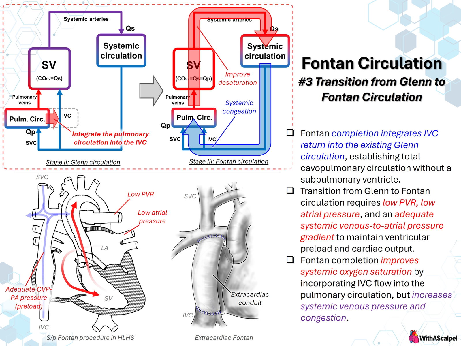
Fontan completion converts the bidirectional Glenn circulation into a total cavopulmonary connection (TCPC) by directing inferior vena caval (IVC) return to the pulmonary arteries. The superior vena cava (SVC) is already connected to the pulmonary circulation after the Glenn procedure. Following Fontan completion, essentially all systemic venous blood traverses the lungs without an intervening subpulmonary ventricle [1,2].
The transition has two fundamental and inseparable effects: systemic oxygen saturation improves, because desaturated IVC blood no longer enters the systemic ventricle directly, while systemic venous pressure increases, because IVC blood must now cross the pulmonary circulation under passive-flow conditions. Successful Fontan physiology requires pulmonary blood flow sufficient to maintain systemic ventricular preload and cardiac output without unacceptable systemic venous congestion [1,2].
The Fontan is therefore more than an anatomical operation. It replaces an incompletely separated circulation with a series circulation whose performance depends critically on pulmonary vascular resistance (PVR), pulmonary venous atrial pressure, and resistance within the cavopulmonary pathway.
2. Glenn Circulation Before Fontan Completion
After the bidirectional Glenn, SVC blood passes directly to the pulmonary arteries. IVC blood continues to return to the atrium and single ventricle, where it mixes with pulmonary venous blood. Consequently, Glenn physiology remains cyanotic, even when pulmonary venous oxygenation is normal.
The Glenn circulation is also a stage of ventricular volume unloading. SVC return no longer passes through the ventricle before reaching the pulmonary arteries, decreasing the ventricular volume burden relative to an earlier parallel circulation. Staged Glenn palliation can improve subsequent ventriculoarterial coupling and mechanical efficiency at Fontan completion, although these findings arise principally from observational and physiological studies [3].
The remaining systemic desaturation depends on the balance between SVC and IVC flow, additional antegrade pulmonary blood flow, pulmonary venous saturation, collateral vessels, and intracardiac streaming. It cannot be interpreted solely as an index of pulmonary vascular adequacy. Importantly, the Glenn pulmonary circuit only needs to accommodate SVC return; after Fontan completion it must accommodate both caval streams.
3. Incorporation of IVC Return
Fontan completion redirects IVC flow through an extracardiac conduit or an intra-atrial lateral tunnel to the pulmonary arteries. An extracardiac Fontan creates an external pathway between the IVC and pulmonary arteries, whereas a lateral-tunnel Fontan uses an intracardiac baffle. The two techniques differ anatomically but share the same physiological purpose: to incorporate lower-body systemic venous return into the cavopulmonary circuit.
For an ideal, nonfenestrated Fontan without major collaterals or residual shunts:
In this series circulation, systemic ventricular output must equal the effective pulmonary flow that returns through the pulmonary veins. This contrasts with earlier parallel single-ventricle physiology, in which the ventricle may eject the combined systemic and recirculating pulmonary flows.
The crucial consequence is that IVC return no longer provides direct ventricular filling. It must first traverse the passive pulmonary circulation. Pulmonary venous return becomes the principal source of ventricular preload. The ventricle cannot compensate for impaired transpulmonary flow merely by increasing contractility [2,4].
4. Pressure Gradient and Pulmonary Flow
Because the Fontan circuit has no subpulmonary ventricle, the pressure gradient between the systemic venous compartment and the pulmonary venous atrium sustains pulmonary blood flow. A simplified relationship is:
Here, PFontan is the systemic venous/Fontan pathway pressure, PLA is the pulmonary venous atrial pressure, RTCPC represents pathway resistance, and PVR represents resistance within the pulmonary vasculature. The equation is a conceptual steady-flow approximation; actual Fontan flow is influenced by respiratory fluctuations, vessel compliance, energy losses, and flow distribution.
In an unobstructed Fontan, central venous and pulmonary artery pressures are usually similar. Thus, a “CVP-to-PA pressure gradient” is not the main driving gradient. The clinically relevant pressure difference is from the Fontan/pulmonary arterial compartment to the pulmonary venous atrium. Pulmonary arterial obstruction can add a separate pressure drop before blood even enters the distal pulmonary vascular bed.
A rise in PVR or left atrial pressure either reduces flow at a given Fontan pressure or requires a further increase in systemic venous pressure to maintain the same flow. Both mechanisms compromise the narrow operating range of Fontan physiology [2,4,5].
5. Why Low PVR Is Essential
In normal biventricular physiology, the right ventricle can increase pulmonary arterial pressure when resistance rises. The Fontan has no such pump. Even a modest increase in PVR may therefore compromise pulmonary flow, ventricular preload, and systemic output [2,5,6].
For this reason, conventional pulmonary hypertension thresholds are not directly applicable to Fontan physiology. A patient may have a relatively low absolute pulmonary artery pressure while a small increase in PVR causes clinically important flow limitation. Pulmonary vascular health is determined not simply by pressure but by the relation among resistance, flow, and the pressure available to drive that flow [6].
In a catheterization cohort of 261 adult Fontan patients, elevated pulmonary vascular resistance index combined with reduced cardiac index was associated with higher risk of Fontan failure [7]. A 2026 Pediatric Cardiac Care Consortium study involving 1,175 patients also found that higher pre-Fontan mean pulmonary artery pressure predicted in-hospital failure and long-term death or transplant, with a continuous association rather than an obvious universal cutoff [8]. These observations support a comprehensive hemodynamic assessment rather than reliance on one pressure threshold.
6. Why Low Atrial Pressure Is Equally Important
A low-resistance pulmonary vascular bed is insufficient if downstream filling pressure is elevated. The pulmonary venous atrium must provide a low-pressure receiving compartment so that blood can traverse the lungs.
For example, if Fontan pressure is 14 mmHg and atrial pressure increases from 5 to 9 mmHg, the effective pressure gradient falls from 9 to 5 mmHg. Unless resistance decreases, pulmonary flow is reduced. To preserve flow, systemic venous pressure would have to rise further.
Ventricular diastolic dysfunction, reduced ventricular compliance, significant atrioventricular valve regurgitation, and pulmonary venous obstruction can all increase downstream pressures. These abnormalities may cause low cardiac output and elevated Fontan pressure even when the conduit and branch pulmonary arteries appear widely patent.
Pre-Fontan evaluation must therefore examine the entire circuit: PVR, mean pulmonary artery pressure, pulmonary venous return, ventricular filling pressure, systolic and diastolic ventricular function, atrioventricular valve competence, and systemic outflow. Historical and contemporary series emphasize the importance of pulmonary and ventricular filling hemodynamics in determining Fontan outcomes [1,9].
7. The Necessary Increase in Systemic Venous Pressure
After completion, IVC flow can no longer return directly to the systemic ventricle. To generate pulmonary flow, systemic venous pressure must exceed downstream atrial pressure sufficiently to overcome resistance across the TCPC and pulmonary vascular bed. This produces chronically elevated Fontan pressure.
This is an intrinsic feature of Fontan physiology, not simply a postoperative complication. However, there is a crucial distinction between the venous pressure necessary to sustain flow and excessive venous pressure that produces congestion.
If central venous pressure is insufficient, pulmonary flow and ventricular preload fall. Conversely, raising venous pressure through additional intravascular volume may fail to improve cardiac output once the cavopulmonary circuit becomes the limiting resistance. The consequence may instead be increased hepatic congestion, lymphatic hypertension, effusions, ascites, and peripheral edema.
The optimal operating condition is therefore adequate transpulmonary flow at the lowest feasible systemic venous pressure. The characteristic combination of upstream venous congestion and downstream preload deprivation has been described as a fundamental paradox of the Fontan circulation [2,4].
8. Improved Saturation Versus Venous Congestion
Fontan completion usually increases systemic arterial oxygen saturation because IVC blood is oxygenated before it returns to the single ventricle. This reduces the chronic systemic venous admixture characteristic of the Glenn stage.
However, improved arterial saturation must not be confused with improved oxygen delivery. Systemic oxygen delivery depends on arterial oxygen content and cardiac output. A patient with nearly normal arterial saturation but insufficient transpulmonary flow may have low systemic oxygen delivery and inadequate organ perfusion.
Fenestration illustrates this tradeoff. A Fontan fenestration provides a communication between the high-pressure cavopulmonary pathway and the lower-pressure atrium. Right-to-left flow may lower Fontan pressure and augment ventricular filling at the expense of systemic desaturation and potential paradoxical embolic risk. Early modified Fontan experience demonstrated this concept of controlled systemic venous decompression [10,11].
The physiological balance is therefore not simply improved saturation versus residual cyanosis. It is better oxygenation and separation of flows versus the pressure and resistance burden imposed on the systemic venous circulation.
9. Surgical Geometry and Completion Assessment
Because pulmonary flow depends on a limited available pressure gradient, unnecessary resistance within the Fontan pathway should be avoided. Conduit narrowing, anastomotic distortion, kinking, branch pulmonary artery stenosis, compression, and adverse stream geometry can all increase energy loss.
Computational and physiological modeling has shown that resistance within the TCPC can substantially affect cardiac output and exercise reserve, reinforcing the importance of unobstructed conduit-to-pulmonary artery flow [5].
An extracardiac Fontan requires attention to conduit length and diameter, alignment with the pulmonary artery confluence, and freedom from tension or torsion. The distal anastomosis must distribute flow without creating avoidable branch PA obstruction. In patients with complex pulmonary arterial anatomy, preoperative imaging and individualized reconstruction may be necessary.
Completion assessment should verify conduit patency, SVC and IVC pathway pressures, branch pulmonary artery flow, unobstructed pulmonary venous return, ventricular filling pressure, atrioventricular valve competence, and ventricular function. Where relevant, the degree of fenestration flow and residual shunting should be correlated with oxygen saturation and cardiac output.
10. Early Postoperative Physiology
Immediately following Fontan completion, small changes in PVR, intrathoracic pressure, circulating volume, ventricular compliance, or conduit resistance may produce disproportionately large changes in cardiac output.
Important management principles include:
- Maintain low PVR. Avoid hypoxemia, substantial hypercarbia, acidosis, atelectasis, and excessive airway pressures.
- Preserve adequate effective preload. Hypovolemia impairs cavopulmonary flow, but indiscriminate volume loading may worsen venous congestion without increasing ventricular filling.
- Minimize downstream pressure. Ventricular diastolic impairment, ventricular dysfunction, and atrioventricular valve regurgitation reduce the effective transpulmonary gradient.
- Maintain unobstructed pulmonary flow. Branch pulmonary artery stenosis, Fontan pathway obstruction, thrombus, and pulmonary venous obstruction require timely recognition.
- Optimize respiratory interaction. Favorable spontaneous respiratory mechanics can facilitate venous return and transpulmonary flow, whereas high intrathoracic pressure may impair both.
- Preserve effective rhythm and atrioventricular synchrony. Tachyarrhythmia and loss of atrial contribution can further compromise ventricular filling.
A high Fontan pressure accompanied by low output should prompt a structured search for elevated PVR, mechanical obstruction, increased atrial pressure, impaired ventricular relaxation, important valve regurgitation, or adverse ventilatory interaction rather than reflexively treating the pressure value with additional volume.
Key Clinical Principles
- Fontan completion incorporates IVC return into the existing Glenn pathway to establish a total cavopulmonary connection.
- Systemic oxygen saturation improves, but this benefit comes with a necessary increase in systemic venous pressure.
- Pulmonary flow determines ventricular preload when no subpulmonary ventricle is present.
- Low PVR and low atrial pressure are equally important for maintaining the transpulmonary driving gradient.
- CVP and PA pressure are normally similar in an unobstructed Fontan; the principal gradient is toward the pulmonary venous atrium.
- High oxygen saturation does not guarantee sufficient cardiac output or adequate systemic oxygen delivery.
- Conduit geometry and branch PA patency matter because small additional resistive losses can have major physiological consequences.
- Optimal Fontan physiology requires adequate flow with the lowest sustainable venous pressure, rather than maximal volume loading or maximal oxygen saturation alone.
References
- Rychik J, Atz AM, Celermajer DS, et al. Evaluation and Management of the Child and Adult With Fontan Circulation: A Scientific Statement From the American Heart Association. Circulation. 2019. DOI: 10.1161/CIR.0000000000000696. PMID: 31256636.
- Gewillig M, Brown SC. The Fontan circulation after 45 years: update in physiology. Heart. 2016. DOI: 10.1136/heartjnl-2015-307467. PMID: 27220691.
- Tanoue Y, Sese A, Ueno Y, Joh K, Hijii T. Bidirectional Glenn Procedure Improves the Mechanical Efficiency of a Total Cavopulmonary Connection in High-Risk Fontan Candidates. Circulation. 2001. DOI: 10.1161/01.CIR.103.17.2176. PMID: 11331259.
- Ridderbos FS, Hoendermis ES, Berger RMF, van Melle JP. The Janus-faced Fontan circulation: unravelling its elusive pathophysiology. European Journal of Heart Failure. 2019. DOI: 10.1002/ejhf.1415. PMID: 30730092.
- Sundareswaran KS, Pekkan K, Dasi LP, et al. The total cavopulmonary connection resistance: a significant impact on single ventricle hemodynamics at rest and exercise. American Journal of Physiology-Heart and Circulatory Physiology. 2008. DOI: 10.1152/ajpheart.00628.2008. PMID: 18931028.
- Becker K, Uebing A, Hansen JH. Pulmonary vascular disease in Fontan circulation—is there a rationale for pulmonary vasodilator therapies? Cardiovascular Diagnosis and Therapy. 2020. DOI: 10.21037/CDT-20-431. PMID: 34527537.
- Egbe AC, Connolly HM, Miranda WR, et al. Hemodynamics of Fontan Failure: The Role of Pulmonary Vascular Disease. Circulation: Heart Failure. 2017. DOI: 10.1161/CIRCHEARTFAILURE.117.004515. PMID: 29246897.
- Suthar D, Yang YX, Beshish AG, et al. Association of pre-Fontan hemodynamics with long-term outcomes after Fontan palliation: A study from the Pediatric Cardiac Care Consortium. Circulation. 2026. DOI: 10.1161/CIRCULATIONAHA.125.076880. PMID: 41568447.
- Kido T, Burri M, Mayr B, et al. Impacts of stage 1 palliation and pre-Glenn pulmonary artery pressure on long-term outcomes after Fontan operation. European Journal of Cardio-Thoracic Surgery. 2021. DOI: 10.1093/ejcts/ezab079. PMID: 33764447.
- Laks H. The partial Fontan procedure. A new concept and its clinical application. Circulation. 1990. DOI: 10.1161/01.CIR.82.5.1866. PMID: 2225386.
- Laks H, Ardehali A, Grant PW, et al. Modification of the Fontan procedure: superior vena cava to left pulmonary artery connection and inferior vena cava to right pulmonary artery connection with adjustable atrial septal defect. Circulation. 1995. DOI: 10.1161/01.CIR.91.12.2943. PMID: 7796504.