1. Overview
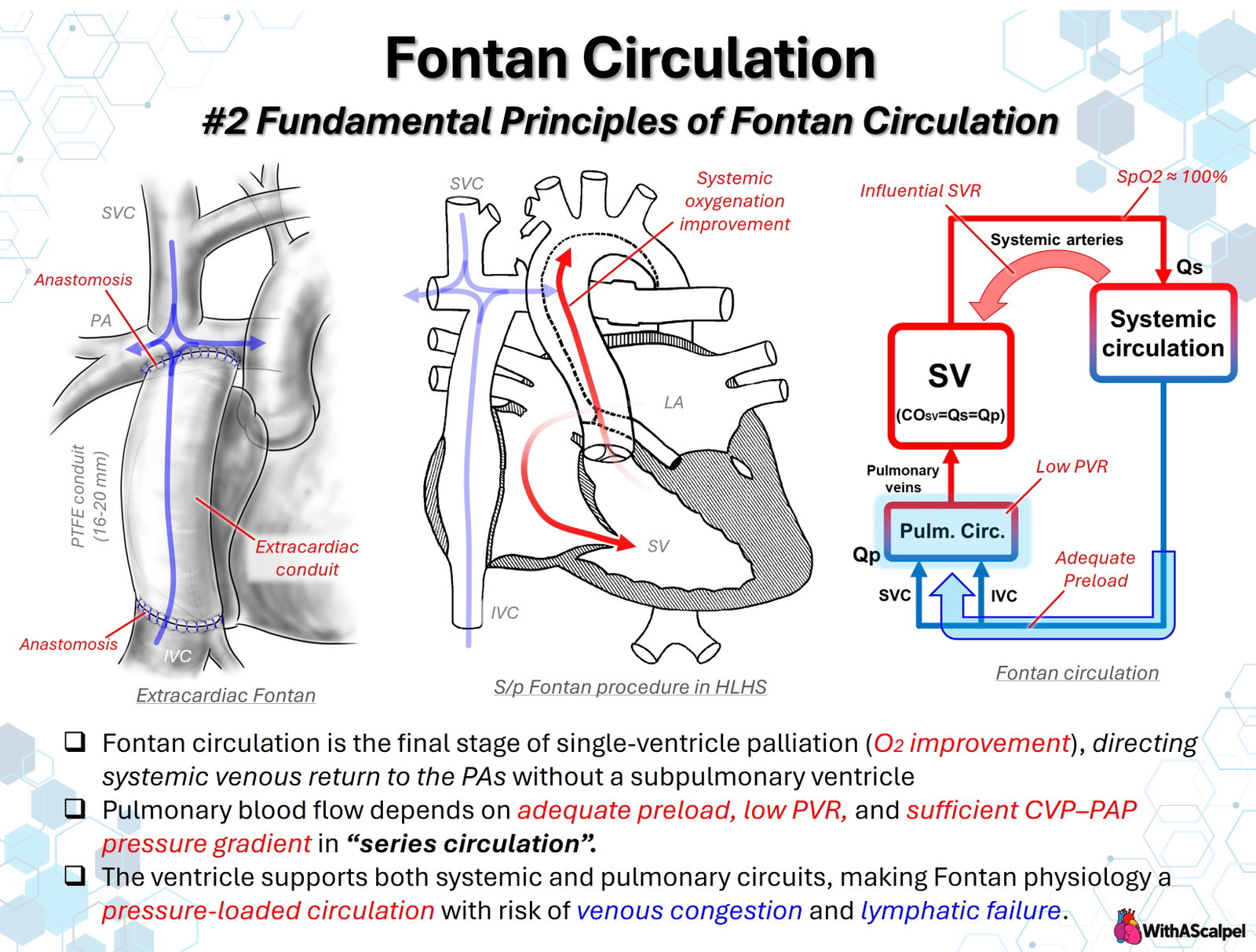
Fontan circulation is the definitive palliative circulation for patients with a functional single ventricle when a durable biventricular repair is not feasible. Its defining feature is not a particular conduit or tunnel but a hemodynamic arrangement: systemic venous return is directed to the pulmonary arteries without an intervening subpulmonary ventricle. The pulmonary and systemic vascular beds are therefore connected in series, and the single functional ventricle serves as the systemic pump [1–4].
This arrangement provides two major advantages compared with the pre-Fontan parallel circulation. First, systemic and pulmonary venous blood are largely separated, allowing systemic arterial oxygen saturation to approach the normal range. Second, the chronic ventricular volume load produced by recirculation of pulmonary blood is markedly reduced. These benefits come at an unavoidable physiological cost: pulmonary blood flow must traverse the lungs without a right-sided pumping chamber. Fontan circulation therefore depends on chronically elevated systemic venous pressure, low pulmonary vascular resistance, and sufficient ventricular filling downstream of the pulmonary circuit [1–4].
The Fontan operation therefore creates a new limiting element—the cavopulmonary-pulmonary vascular pathway. The systemic ventricle remains essential but cannot directly force blood through this upstream bottleneck [3,4].
2. Conversion to a Series Circulation
Before Fontan completion, pulmonary and systemic blood flows are not fully separated. The bidirectional Glenn directs superior vena caval return to the pulmonary arteries, but inferior vena caval blood still returns to the heart.
Fontan completion incorporates the inferior vena caval pathway, usually through an extracardiac conduit or lateral tunnel, so that essentially all systemic venous return is directed to the pulmonary arteries. In an ideal nonfenestrated Fontan with no important collateral shunts:
Qp ≈ Qs ≈ systemic ventricular output
This equality reflects the series arrangement: blood leaving the systemic ventricle must traverse the systemic vascular bed, Fontan pathway, pulmonary circulation, and pulmonary veins before returning to the ventricle [1–3].
This does not mean that the ventricle actively pumps both circuits in the same way as two ventricles in a normal heart. Rather, the single systemic ventricle provides the energy for the entire closed loop, while the pulmonary circulation receives no direct systolic pressure boost from a subpulmonary pump. Residual hydraulic energy after passage through the systemic circulation, together with elevated systemic venous pressure and favorable intrathoracic mechanics, must be sufficient to maintain pulmonary flow.
3. The Fontan Paradox: High Venous Pressure and Low Pulmonary Artery Pressure
A normal biventricular circulation uses the right ventricle to maintain a low systemic venous pressure while generating enough pulmonary arterial pressure to overcome pulmonary vascular resistance. The Fontan circulation reverses this relationship. The systemic veins and Fontan pathway must operate at a higher pressure, whereas pulmonary artery pressure remains relatively low because there is no ventricular chamber directly upstream of the pulmonary arteries [1,3].
This produces the characteristic Fontan paradox:
upstream systemic venous hypertension + downstream preload limitation
The cavopulmonary circuit behaves as a hydrodynamic bottleneck. If resistance within the Fontan pathway or pulmonary vasculature increases, blood accumulates upstream as venous congestion, while flow downstream falls. The ventricle cannot fully compensate by increasing contractility because the primary limitation is inadequate filling rather than inadequate ejection [2–4].
Modern hydrodynamic models emphasize that control of cardiac output shifts toward the Fontan “neoportal” system—the vascular pathway connecting systemic and pulmonary capillary beds without an intervening pump [4]. Consequently, increasing contractility alone may have little effect when excessive resistance or elevated downstream filling pressure is dominant.
4. The Pressure Gradient That Drives Pulmonary Blood Flow
Pulmonary blood flow follows the same basic relationship as flow through any resistive circuit:
Flow ≈ pressure gradient / resistance
For the Fontan circulation, a useful approximation is:
Qp ≈ (Fontan pressure − left atrial pressure) / (Fontan pathway resistance + pulmonary vascular resistance)
The clinically relevant driving pressure is therefore the gradient from the systemic venous/Fontan compartment through the pulmonary circulation to the pulmonary venous atrial compartment. In an unobstructed total cavopulmonary connection, Fontan pressure and pulmonary artery pressure are usually close to one another. Thus, describing pulmonary flow as dependent on a “CVP-to-PA pressure gradient” is incomplete; the more important gradient is the transpulmonary or transpulmonary-circuit gradient from Fontan pressure toward left atrial pressure [2,5].
Several abnormalities can reduce this gradient or increase resistance: conduit or pathway narrowing, branch pulmonary artery stenosis, pulmonary vascular disease, pulmonary venous obstruction, elevated left atrial pressure, high ventricular end-diastolic pressure, and excessive intrathoracic pressure. Each lesion may produce the same final physiology—reduced pulmonary flow, reduced ventricular preload, and decreased systemic cardiac output.
5. Low Pulmonary Vascular Resistance Is the Central Requirement
Because there is no subpulmonary ventricle, the Fontan circulation has very little pressure reserve with which to overcome an increase in pulmonary vascular resistance (PVR). Even modest increases in PVR may substantially reduce pulmonary blood flow or require a higher systemic venous pressure to preserve flow [3,5,6].
This is why low PVR is a fundamental requirement before and after Fontan completion. The importance is not simply that elevated PVR raises pulmonary artery pressure; in fact, pulmonary artery pressure may remain relatively low in Fontan patients despite clinically important pulmonary vascular disease. The more important consequence is that increased resistance reduces transpulmonary flow and ventricular preload [5].
Observational catheterization data support this physiological model. In adult Fontan patients, the combination of elevated pulmonary vascular resistance index and low cardiac index has been associated with increased risk of Fontan failure [6]. These data should not be interpreted as establishing a single universal threshold for intervention, but they reinforce the principle that even relatively small abnormalities in the pulmonary vascular bed may be clinically important in a circulation without a subpulmonary pump.
6. Preload Is Necessary—but Preload Reserve Is Limited
The systemic ventricle can eject only the blood that reaches it through the lungs. Consequently, pulmonary blood flow is ventricular preload in Fontan circulation. This is one of the most important differences from conventional biventricular heart failure physiology [2–4].
Adequate circulating blood volume is therefore necessary. Hypovolemia lowers mean systemic filling pressure, reduces Fontan flow, and can rapidly decrease cardiac output. However, volume administration has a ceiling. Once the pulmonary circuit becomes the limiting resistance, additional intravascular volume may increase Fontan pressure without meaningfully increasing flow. The result is more systemic venous congestion, tissue edema, hepatic congestion, and lymphatic load rather than improved ventricular filling.
The concept of “adequate preload” should therefore not be translated into indiscriminate volume loading. The goal is sufficient venous filling pressure to sustain transpulmonary flow at the lowest feasible systemic venous pressure. A patient with high Fontan pressure and low cardiac output may need relief of obstruction, reduction of PVR, improvement in ventricular diastolic pressure, or creation/reopening of a fenestration rather than additional volume.
7. Systemic Vascular Resistance and Ventricular Afterload
Systemic vascular resistance (SVR) remains an important determinant of systemic ventricular afterload. Excessive vasoconstriction can increase ventricular work and limit stroke volume, whereas appropriate systemic vasodilation may reduce afterload. However, SVR should not be considered the primary regulator of Fontan output when pulmonary flow is the dominant bottleneck [2–4].
Lowering SVR cannot compensate for severe PVR elevation, Fontan pathway obstruction, or high ventricular filling pressure. Excessive vasodilation may reduce arterial pressure without generating preload, whereas marked vasoconstriction can worsen ventricular afterload. SVR should therefore be interpreted with Fontan pressure, atrial pressure, ventricular function, and systemic perfusion.
8. Respiration and Cardiopulmonary Interaction
Respiration has an unusually important influence on Fontan flow. Spontaneous inspiration lowers intrathoracic pressure, which can facilitate venous return and cavopulmonary flow. Doppler studies have demonstrated substantial respiratory modulation of infradiaphragmatic and hepatic venous flow in patients with total cavopulmonary connections [7].
Positive-pressure ventilation can have the opposite effect by increasing intrathoracic pressure, reducing the pressure gradient for venous return, and potentially increasing PVR. This is one reason early extubation and spontaneous breathing are often favorable after uncomplicated Fontan completion. Nevertheless, the principle should not be oversimplified: adequate oxygenation, lung recruitment, correction of atelectasis, and control of hypercarbia and acidosis are also essential because hypoxia and poor lung mechanics can increase PVR. The optimal strategy is therefore to maintain well-expanded lungs with the lowest effective mean airway pressure while avoiding hypoxemia, hypercapnia, and acidosis.
9. Oxygen Saturation: Important, but Not the Primary Measure of Fontan Performance
A successful nonfenestrated Fontan markedly reduces intracardiac mixing, so systemic arterial saturation may approach 100%. However, normal saturation is not synonymous with normal Fontan physiology. Patients may have excellent oxygen saturation despite low cardiac output if pulmonary flow is limited but all blood reaching the systemic ventricle is fully oxygenated.
Conversely, mild desaturation may be compatible with acceptable systemic oxygen delivery when a fenestration intentionally permits right-to-left shunting to preserve preload and cardiac output. Other causes of desaturation include systemic-to-pulmonary venous collaterals, pulmonary arteriovenous malformations, residual intracardiac shunts, pulmonary venous desaturation, and lung disease [4].
The relevant endpoint is therefore systemic oxygen delivery, which depends on both arterial oxygen content and cardiac output. Near-normal saturation can coexist with inadequate systemic flow.
10. Venous Congestion and Lymphatic Failure
The elevated systemic venous pressure required to drive Fontan flow is not a benign adaptation. Chronic venous hypertension affects the liver, gastrointestinal tract, kidneys, peripheral tissues, and lymphatic circulation. The lymphatic system is particularly vulnerable because elevated venous pressure both increases lymph production and raises the pressure against which lymph must drain [4,8].
Direct imaging and physiological studies have demonstrated abnormal lymphatic morphology and impaired lymphatic pumping in Fontan patients [8]. In susceptible patients, this physiology contributes to protein-losing enteropathy, plastic bronchitis, chylous effusions, and other manifestations of lymphatic failure [8,9].
These complications illustrate the central tradeoff: venous pressure is required to sustain pulmonary flow, yet the same pressure promotes end-organ and lymphatic injury.
11. Practical Hemodynamic Interpretation
When a Fontan patient develops low cardiac output, rising venous pressure, effusions, or worsening exercise tolerance, the circulation should be analyzed systematically from upstream to downstream. Important questions include whether circulating volume is adequate, whether the Fontan pathway and branch pulmonary arteries are unobstructed, whether PVR has increased, whether pulmonary venous or left atrial pressure is elevated, and whether ventricular systolic or diastolic dysfunction is limiting filling.
Modern Fontan physiology emphasizes that the ventricle may become a secondary limitation after the cavopulmonary circuit has already restricted preload [3,4,10]. Therapy should target the dominant bottleneck: anatomical obstruction, reversible pulmonary vascular tone, adverse lung mechanics, elevated filling pressure, rhythm disturbance, or—when appropriate—the need for controlled decompression.
Key Clinical Principles
- Fontan completion converts single-ventricle physiology into a series circulation in which systemic venous return reaches the pulmonary arteries without a subpulmonary ventricle.
- In an ideal nonfenestrated Fontan, Qp ≈ Qs, but collateral flow and fenestration can alter this relationship.
- Pulmonary blood flow determines ventricular preload, making low PVR and low downstream filling pressure fundamental to cardiac output.
- The relevant driving gradient is from the Fontan/systemic venous compartment toward the pulmonary venous atrium, not simply from CVP to the pulmonary artery.
- High systemic venous pressure and restricted preload coexist by design; this is the central physiological tradeoff of Fontan circulation.
- Adequate intravascular volume is necessary, but excessive volume may increase venous congestion without increasing flow.
- SVR influences ventricular afterload but cannot overcome a fixed cavopulmonary or pulmonary vascular bottleneck.
- Respiration matters: spontaneous negative intrathoracic pressure generally supports Fontan flow, whereas excessive positive-pressure ventilation may impair it.
- SpO₂ alone does not define Fontan success; systemic oxygen delivery depends on both oxygen content and cardiac output.
- Venous and lymphatic complications are intrinsic consequences of the pressure-loaded systemic venous circulation, not merely late surgical complications.
References
- Gewillig M. The Fontan circulation. Heart. 2005. DOI: 10.1136/hrt.2004.051789. PMID: 15894794.
- Gewillig M, Brown SC, Eyskens B, et al. The Fontan circulation: who controls cardiac output? Interactive Cardiovascular and Thoracic Surgery. 2010. DOI: 10.1510/icvts.2009.218594. PMID: 19995891.
- Gewillig M, Brown SC. The Fontan circulation after 45 years: update in physiology. Heart. 2016. DOI: 10.1136/heartjnl-2015-307467. PMID: 27220691.
- Rychik J, Atz AM, Celermajer DS, et al. Evaluation and management of the child and adult with Fontan circulation: a scientific statement from the American Heart Association. Circulation. 2019. DOI: 10.1161/CIR.0000000000000696. PMID: 31256636.
- Becker K, Uebing A, Hansen JH. Pulmonary vascular disease in Fontan circulation—is there a rationale for pulmonary vasodilator therapies? Cardiovascular Diagnosis and Therapy. 2021. DOI: 10.21037/cdt-20-431. PMID: 34527537.
- Egbe AC, Connolly HM, Miranda WR, et al. Hemodynamics of Fontan failure: the role of pulmonary vascular disease. Circulation: Heart Failure. 2017. DOI: 10.1161/CIRCHEARTFAILURE.117.004515. PMID: 29246897.
- Hsia TY, Khambadkone S, Redington AN, Migliavacca F, Deanfield JE, de Leval MR. Effects of respiration and gravity on infradiaphragmatic venous flow in normal and Fontan patients. Circulation. 2000. DOI: 10.1161/01.CIR.102.SUPPL_3.III-148. PMID: 11082378.
- Mohanakumar S, Telinius N, Kelly B, et al. Morphology and function of the lymphatic vasculature in patients with a Fontan circulation. Circulation: Cardiovascular Imaging. 2019. DOI: 10.1161/CIRCIMAGING.118.008074. PMID: 30943769.
- Sharma VJ, Iyengar AJ, Zannino D, et al. Protein-losing enteropathy and plastic bronchitis after the Fontan procedure. Journal of Thoracic and Cardiovascular Surgery. 2020. DOI: 10.1016/j.jtcvs.2020.07.107. PMID: 32928546.
- Gewillig M, Salaets T, Van de Bruaene A, Van den Eynde J, Brown SC. How cardiac output is controlled in a Fontan circulation: an update. Interdisciplinary Cardiovascular and Thoracic Surgery. 2025. DOI: 10.1093/icvts/ivae183. PMID: 39906971.