1. Definition and Clinical Significance
Fontan failure occurs when total cavopulmonary circulation cannot sustain adequate systemic blood flow without excessive systemic venous pressure or clinically consequential congestion. In the early postoperative period, it may present as low cardiac output, escalating vasoactive support, persistent lactic acidosis, elevated Fontan pressure, refractory pleural drainage, or multiorgan dysfunction. Early Fontan failure is often defined in research by major events within 30 days, although the most urgent rescue decisions commonly arise in the operating room or during the first postoperative days [1,2].
Unlike a biventricular circulation, the Fontan pathway has no subpulmonary pump. Pulmonary blood flow depends on the pressure difference between the systemic veins and pulmonary venous atrium, opposed by resistance throughout the cavopulmonary pathway and pulmonary vascular bed. Increased pulmonary vascular resistance (PVR), branch pulmonary artery obstruction, elevated ventricular filling pressure, and atrioventricular (AV) valve regurgitation can therefore produce both upstream venous hypertension and downstream ventricular preload deprivation. These mechanisms are distinct, frequently coexist, and determine which rescue intervention is appropriate [1].
The therapeutic objective is not simply to lower a measured central venous pressure. It is to restore effective pulmonary transit and systemic oxygen delivery while preventing progressive venous and end-organ injury.
2. Rapid Hemodynamic and Anatomic Assessment
Early deterioration requires parallel resuscitation and identification of its mechanism. Clinical monitoring should integrate arterial pressure, lactate trends, mixed or central venous oxygen saturation, cerebral and somatic near-infrared spectroscopy, urine output, chest drainage, and rhythm. Simultaneous measurement of Fontan or superior vena caval pressure and pulmonary venous atrial pressure helps distinguish impaired transpulmonary passage from elevated downstream filling pressure.
A high Fontan pressure with low atrial pressure suggests important resistance between the systemic veins and left atrium, whether from PVR, pulmonary arterial obstruction, pulmonary venous disease, or mechanical distortion. Elevation of both venous and atrial pressures may indicate ventricular diastolic dysfunction, impaired ventricular ejection, significant AV valve regurgitation, or combinations of these abnormalities. These patterns guide investigation; neither pressure difference alone nor an arbitrary cutoff defines the treatment.
Echocardiography should reassess ventricular systolic and diastolic function, AV valve competence, pulmonary venous return, residual atrial restriction, and any accessible Fontan pathway. Angiography, catheterization, computed tomography, or operative inspection may be necessary when an anatomic obstruction is suspected or noninvasive imaging is inadequate. Small pressure gradients can still matter within a low-energy cavopulmonary circuit. Early failure should never be attributed to intrinsically unfavorable Fontan physiology without first excluding a correctable residual lesion [2].
3. Pulmonary Vascular Resistance Reduction and Intensive Care Rescue
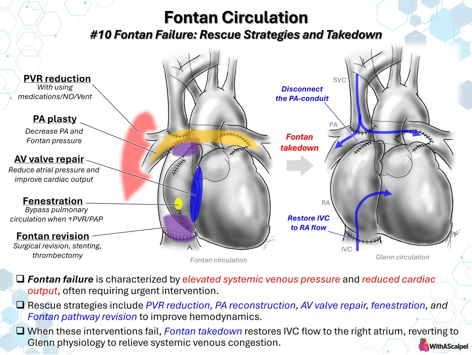
Reducing pulmonary vascular impedance is often the first physiologic intervention. Hypoxemia, hypercarbia, acidosis, hypothermia, atelectasis, pulmonary edema, and excessive airway pressure may raise resistance or impede venous return. Management therefore emphasizes adequate oxygenation, temperature and acid–base correction, lung recruitment without overdistension, and ventilation that limits unnecessary mean intrathoracic pressure.
Ventilation must be individualized. Spontaneous breathing may facilitate cavopulmonary flow by generating negative intrathoracic pressure, but premature extubation in a child with severe ventricular dysfunction, increased work of breathing, or inadequate gas exchange may worsen systemic oxygen delivery. Positive-pressure ventilation remains necessary in many unstable patients; tidal volume, positive end-expiratory pressure, and recruitment should balance pulmonary mechanics against adverse effects on venous return.
Inhaled nitric oxide offers selective pulmonary vasodilation and has demonstrated acute hemodynamic benefit in selected patients after Fontan surgery. Its response depends on the contribution of reactive pulmonary vasoconstriction to the patient's physiology. Published postoperative series support reductions in venous pressure or transpulmonary gradient, but do not establish a universal survival benefit [3,4]. Phosphodiesterase-5 inhibition and other pulmonary vasodilators may be considered selectively, with attention to systemic blood pressure, evidence of pulmonary vascular responsiveness, and drug-specific effects.
Volume administration requires particular restraint. Additional preload may temporarily support output, but when systemic venous pressure is already markedly elevated, excessive fluid increases hepatic and lymphatic congestion without necessarily augmenting ventricular filling. Inotropes, afterload management, rhythm optimization, and treatment of myocardial ischemia should be tailored to the systemic ventricle and AV valve.
4. Correctable Lesions and Targeted Rescue Procedures
4.1. Pulmonary Artery Reconstruction
Branch pulmonary artery stenosis, distortion near the superior cavopulmonary anastomosis, or narrowing of the central confluence increases the resistance against which the entire Fontan circuit must operate. Treatment may involve surgical patch augmentation, revision of an anastomosis, balloon dilation, or stent implantation. The objective is unobstructed, well-distributed pulmonary blood flow, rather than simply a larger angiographic diameter.
Pulmonary artery plasty should address the full length of the lesion and avoid introducing new angulation, kinking, or excessive patch redundancy. Catheter interventions can be effective for discrete stenoses, although published stent cohorts often include late Fontan obstruction rather than immediate postoperative rescue [5]. Residual branch obstruction should be reconsidered if venous hypertension persists despite apparently satisfactory conduit reconstruction.
4.2. Atrioventricular Valve Repair
Moderate or severe systemic AV valve regurgitation increases atrial pressure, worsens ventricular volume loading, and reduces the pressure gradient available for passive pulmonary flow. A repairable lesion may warrant annuloplasty, leaflet or commissural reconstruction, or correction of tethering and coaptation defects. The technical goal is durable competence without creating inflow stenosis.
The timing of repair must balance the potential hemodynamic gain against additional ischemic time and operative risk in an unstable patient. Results of AV valve reconstruction in single-ventricle patients are heterogeneous, and repair durability is anatomy dependent [6]. In a patient with profound ventricular dysfunction or unreconstructable valve disease, isolated valve repair may not resolve the underlying failure.
4.3. Fenestration
A Fontan fenestration establishes a controlled right-to-left communication from the high-pressure systemic venous pathway to the pulmonary venous atrium. By partially bypassing the pulmonary circulation, it can reduce venous congestion and increase ventricular preload and systemic output. The trade-off is reduced arterial oxygen saturation and potential paradoxical embolization.
A randomized study of 49 patients undergoing Fontan completion demonstrated less pleural drainage and shorter hospitalization with fenestration, although its results do not establish the effectiveness of every postoperative rescue fenestration [7]. In selected patients with an extracardiac conduit, urgent catheter-based fenestration creation or enlargement can be considered; small published series demonstrate technical feasibility and physiologic improvement [8]. The location must permit a controlled communication without impinging on pulmonary venous inflow or adjacent structures.
4.4. Fontan Pathway Revision
Twisting, compression, narrowing, thrombus, unfavorable conduit-to-pulmonary artery geometry, or an obstructed inferior vena caval connection may require catheter intervention or operative revision. Surgical reconstruction should eliminate the obstructing segment while preserving smooth caval entry, pulmonary artery confluence, and unobstructed superior cavopulmonary flow. Thrombectomy or anticoagulation may be required when thrombosis contributes to obstruction; the approach depends on thrombus burden, bleeding risk, and hemodynamic stability.
5. Mechanical Circulatory Support as a Bridge
Venoarterial extracorporeal membrane oxygenation (VA-ECMO) may stabilize a patient with refractory low output, severe hypoxemia, or impending arrest while the team determines whether the circulation is recoverable. It is a bridge to recovery, targeted intervention, takedown, or transplantation, not correction of the underlying anatomical or physiologic problem.
Cannulation must account for separate superior and inferior systemic venous pathways. Inadequate drainage from either territory can perpetuate venous hypertension despite adequate total circuit flow. Central or multiple-site venous drainage, Fontan pathway decompression, and left atrial or ventricular venting may be necessary according to anatomy and ventricular loading. The residual Fontan circuit and pulmonary blood flow must be considered when setting support and assessing myocardial recovery.
A contemporary Extracorporeal Life Support Organization registry analysis reported 48.4% survival to discharge among 281 children supported with ECMO during their Fontan surgical hospitalization. Pre-ECMO arrest and longer time to cannulation were associated with mortality; these observational findings support prompt escalation but do not establish a causal benefit of a specific cannulation threshold [9].
6. Indications and Timing of Fontan Takedown
Fontan takedown converts total cavopulmonary circulation back to an intermediate circulation, usually retaining the bidirectional Glenn connection while restoring inferior vena caval return to the atrium. It is considered when Fontan physiology remains intolerable despite correction of reversible lesions, pulmonary vascular optimization, fenestration, or temporary circulatory support.
Typical circumstances include refractory systemic venous hypertension with progressive organ dysfunction, persistent inadequate ventricular filling and systemic output, severe pulmonary vascular or ventricular limitations, or failure to separate safely from cardiopulmonary bypass after attempted Fontan completion. There is no universally validated pressure, lactate concentration, or duration of ECMO that mandates takedown. Timing requires multidisciplinary judgment about reversibility, the patient's trajectory, and the risk of prolonged ineffective circulation.
Takedown should not necessarily be postponed until every salvage measure has been exhausted. Conversely, an identifiable and rapidly correctable lesion may justify revision rather than abandonment of the Fontan circulation.
7. Operative Strategy for Takedown to Glenn Physiology
The accompanying schematic depicts takedown of an extracardiac conduit Fontan. Two changes define the reconstruction: disconnection of the conduit from the pulmonary artery and restoration of inferior vena caval drainage to the right atrium. The pre-existing superior vena cava–pulmonary artery connection remains functional. Specific reconstruction varies with original anatomy, conduit position, prior caval division, and associated Glenn or pulmonary artery lesions.
A representative sequence is:
- Establish exposure and circulatory control. Plan safe resternotomy or operative re-entry, inspect adhesions and conduit geometry, and obtain cardiopulmonary bypass access appropriate to the aorta and both caval territories. Evaluate the Glenn anastomosis and pulmonary artery confluence before dividing the Fontan circuit.
- Separate the conduit from the pulmonary artery. Control and disconnect the proximal pulmonary arterial anastomosis. Close or reconstruct the pulmonary arterial opening with direct suture or patch augmentation, depending on geometry. The closure must not narrow the confluence, branch pulmonary arteries, or Glenn outflow.
- Restore inferior caval return to the atrium. Divide or remove the relevant extracardiac conduit segment and establish a wide, unobstructed inferior vena cava–right atrial connection. Direct reanastomosis, patch reconstruction, or other tailored techniques may be necessary if the original caval division has produced a gap or distorted orientation.
- Preserve the intermediate circulation. Confirm an unobstructed superior cavopulmonary connection and exclude any unintended communication or mechanical obstruction. Assess whether Glenn-derived pulmonary flow alone is sufficient; selected patients may need additional controlled pulmonary blood flow, but a systemic-to-pulmonary shunt can increase volume load and pulmonary pressure.
- Assess completion physiology. Confirm inferior caval drainage, pulmonary arterial patency, systemic ventricular filling, AV valve function, and oxygen delivery after separation from bypass. Persistent superior vena caval hypertension, severe desaturation, poor ventricular output, or pulmonary artery obstruction warrants immediate reassessment.
For a lateral-tunnel Fontan, takedown requires modification of the intracardiac baffle rather than simple extracardiac conduit division. The relationship of baffle sutures to atrial tissue and venous orifices determines the safest reconstruction. Takedown should not compromise the coronary sinus or hepatic venous drainage.
8. Post-Takedown Physiology and ICU Management
Following takedown, superior vena caval blood continues to reach the lungs through the Glenn connection, whereas inferior caval blood returns to the atrium and contributes to mixed systemic ventricular inflow. The immediate benefit is decompression of the inferior systemic venous territory and restoration of a preload pathway that does not depend on passage of all systemic venous blood through the pulmonary vascular bed.
Lower arterial oxygen saturation is expected and should be interpreted with measured systemic blood flow, hemoglobin concentration, venous oxygen saturation, lactate, and tissue perfusion—not saturation alone. Superior caval pressure may remain elevated if Glenn or pulmonary vascular resistance is excessive, and ventricular dysfunction or AV valve regurgitation may continue to limit recovery.
Early priorities include judicious volume replacement, vasoactive weaning, pulmonary toilet, pleural and lymphatic drainage assessment, rhythm surveillance, and recognition of thrombosis or residual obstruction. Over longer follow-up, persistent cyanosis, restricted exercise capacity, possible pulmonary arteriovenous malformation development from altered hepatic venous effluent distribution, and growth of the pulmonary arteries require surveillance.
9. Outcomes and Future Pathways
In a single-center cohort of 592 primary Fontan operations, Murphy and colleagues identified early failure in 11 patients (1.9%); mortality among those with early failure was 46%. Five underwent takedown, and the reported long-term survival in that very small subgroup was 80% [2]. The estimate should not be generalized to contemporary patients without acknowledging selection and sample size.
In an earlier series, Almond and colleagues reported early survival after takedown in 29 of 53 patients (55%); 19 subsequently achieved another Fontan completion. The findings show that takedown does not necessarily preclude later completion, particularly when reversible abnormalities can be corrected [10].
A separate series of 18 extracardiac Fontan takedowns reported 17 survivors of the immediate post-takedown period, with three later redo Fontan completions [11]. By contrast, the Australia and New Zealand Fontan Registry identified 36 takedowns among 1,540 Fontan patients over four decades, with 44% overall mortality and five successful second Fontan operations. Mortality differed by timing, but confounding by clinical severity prevents interpreting early takedown itself as a proven causal survival intervention [12].
These retrospective cohorts are small, heterogeneous, and span different surgical eras. Their consistent message is that takedown remains a legitimate rescue pathway, potentially allowing prolonged Glenn palliation, later Fontan completion, alternative ventricular reconstruction, or transplantation according to individual anatomy and physiology.
10. Key Surgical Principles
- Diagnose the mechanism before selecting rescue: elevated PVR, obstructed pathway, ventricular dysfunction, and AV valve regurgitation require different interventions.
- Relieve resistance throughout the pulmonary pathway: even localized obstruction may impair passive Fontan flow.
- Use fenestration selectively: venous decompression and improved preload come at the cost of desaturation.
- Escalate support before irreversible injury: ECMO buys time for diagnosis and definitive treatment but cannot substitute for correcting the cause.
- Preserve the Glenn during takedown: restore unobstructed inferior caval return to the atrium without compromising superior cavopulmonary or pulmonary arterial flow.
- Assess oxygen delivery rather than saturation alone: lower saturation may coexist with improved cardiac output and systemic perfusion.
- Maintain a long-term plan: successful takedown can serve as durable intermediate palliation or a bridge to reconstruction or transplantation.
References
- Gewillig M, Brown SC. The Fontan circulation after 45 years: update in physiology. Heart. 2016. DOI: 10.1136/heartjnl-2015-307467. PMID: 27220691.
- Murphy MO, Glatz AC, Goldberg DJ, et al. Management of early Fontan failure: a single-institution experience. European Journal of Cardio-Thoracic Surgery. 2014. DOI: 10.1093/ejcts/ezu022. PMID: 24554071.
- Goldman AP, Delius RE, Deanfield JE, et al. Pharmacological control of pulmonary blood flow with inhaled nitric oxide after the fenestrated Fontan operation. Circulation. 1996. PMID: 8901718.
- Yoshimura N, Yamaguchi M, Oka S, et al. Inhaled Nitric Oxide Therapy After Fontan-Type Operations. Surgery Today. 2004. DOI: 10.1007/s00595-004-2887-1. PMID: 15622461.
- Udink ten Cate FEA, Trieschmann U, Germund I, et al. Stenting the Fontan pathway in paediatric patients with obstructed extracardiac conduits. Heart. 2017. DOI: 10.1136/heartjnl-2016-310511. PMID: 28237969.
- Nakata T, Fujimoto Y, Hirose K, et al. Atrioventricular valve repair in patients with functional single ventricle. Journal of Thoracic and Cardiovascular Surgery. 2010. DOI: 10.1016/j.jtcvs.2010.05.024. PMID: 20584537.
- Lemler MS, Scott WA, Leonard SR, et al. Fenestration Improves Clinical Outcome of the Fontan Procedure: A Prospective, Randomized Study. Circulation. 2002. DOI: 10.1161/HC0202.102237. PMID: 11790702.
- Michel-Behnke I, Luedemann M, Bauer J, et al. Fenestration in Extracardiac Conduits in Children After Modified Fontan Operation by Implantation of Stent Grafts. Pediatric Cardiology. 2004. DOI: 10.1007/s00246-004-0693-6. PMID: 15793660.
- Anton-Martin P, Cavalcante RC, Master L, et al. Extracorporeal membrane oxygenation in children after Fontan operation. Journal of Thoracic and Cardiovascular Surgery. 2026;172:1164–1173.e6. DOI: 10.1016/j.jtcvs.2026.08.002. PMID: 42595216.
- Almond CSD, Mayer JE Jr, Thiagarajan RR, et al. Outcome after Fontan failure and takedown to an intermediate palliative circulation. Annals of Thoracic Surgery. 2007;84:880–887. DOI: 10.1016/j.athoracsur.2007.02.092. PMID: 17720394.
- Trezzi M, Cetrano E, Giannico S, et al. Long-Term Outcomes After Extracardiac Fontan Takedown to an Intermediate Palliative Circulation. Annals of Thoracic Surgery. 2017. DOI: 10.1016/j.athoracsur.2017.05.068. PMID: 28935350.
- Marathe SP, Iyengar AJ, Betts KS, et al. Long-term outcomes following Fontan takedown in Australia and New Zealand. Journal of Thoracic and Cardiovascular Surgery. 2021;161:1126–1135. DOI: 10.1016/j.jtcvs.2020.09.074. PMID: 33131894.